
What are Fungi?
Introduction to fungal infections
Fungal infections in humans can be broadly divided into (i) superficial skin and mucosal infections and (ii) deep or systemic infections. Superficial infections do not necessarily require systemic predisposing factors whereas deep and systemic infections are more commonly seen in immunocompromised or critically ill hosts. Systemic fungal infections are medical emergencies and have high mortality especially if appropriate therapy is delayed.
Examples of fungal infections:
- Skin infection caused by dermatophytes e.g. ringworm, athlete’s foot and fungal nail infections
- Oral and vulvovaginal candidosis caused by C. albicans or other Candida species
- Pulmonary infections e.g. pulmonary aspergillosis, Pneumocystis jirovecii pneumonia, pulmonary histoplasmosis.
- Systemic/deep mycoses e.g. candidaemia, invasive aspergillosis and cryptococcal meningitis
What are Fungi?
Fungi make up one of the five kingdoms of life.
Unlike bacteria, fungi are eukaryotes like humans. This means that their DNA is contained within a nucleus and, in most cases, packed into chromosomes
Unlike animal cells, fungal cells contain a cell-wall which is different from plant and bacterial cell wall.
Unlike plants, fungi do not contain chlorophyll and require pre-formed nutrients for survival.
Fungal cells contain a cell-wall which is typically comprised of glucan, mannan, chitin and chitosan. Detection of some of these components can be used to diagnose systemic fungal infections.
In order to get their nutrients, fungi secrete digestive enzymes, and absorb the released nutrients through the cell wall.
Some fungi are multicellular and some are unicellular.
The number of sets of chromosomes in each fungal cell can vary from the standard 2 (diploid) to 1 (haploid) or many (polyploid). Haploid cells are capable of sexual reproduction leading to new recombinations of genetic material, whereas polyploidy is a quick way for fungal cells to increase the production of essential metabolites by generating multiple copies of the genes encoding for them. These chromosomal variations are employed in the development of antifungal resistance. They can be difficult to detect by standard antifungal susceptibility testing methods as the cells revert back to their standard ploidy quickly in the absence of the drug.
Human pathogenic fungi
There are over 500 species of fungi that have been associated with human disease and approximately 100 of them can cause infection in immunocompetent individuals. Most human fungal infections are endogenous and caused by colonising commensals (mainly yeasts). Others are caused by environmental opportunistic fungi (mainly moulds) that are acquired through inhalation or traumatic implantation. Pathogenic fungi can be broadly divided into 3 main groups:
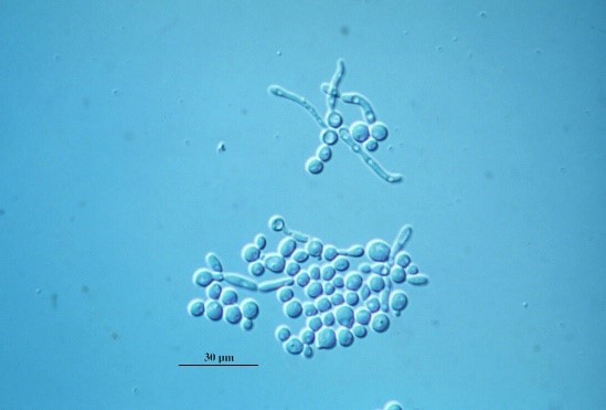
- Yeasts are unicellular fungi, some of which can produce filament-like extensions called pseudohyphae.
- Moulds are multicellular filamentous fungi
- Endemic dimorphic fungi which change their growth pattern depending on the surrounding environment and conditions for growth. They grow as yeasts in human tissues and as moulds in the environment.
Both unicellular and multicellular fungal pathogens form biofilms readily. This is clinically relevant as these multicellular fungal communities are highly resistant to antifungal drugs and cannot be treated without physical disruption or removal of the biofilms.
Fungal biofilms form within hours after introduction on any stable enough surface such as catheters, lines, stents, medical devices or kidney stones. They can also form fungal balls if entrapped into body cavities such as paranasal sinuses or tuberculous cavities .
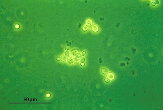
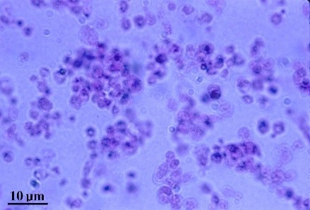
Yeasts
Yeasts are round or ovoid single cells which multiply by budding . The daughter cells will either detach from the mother cell or remain attached and form chains of cells. In tissue, some many yeasts can form filament-like extensions called pseudohyphae which they use for adherence and tissue invasion.
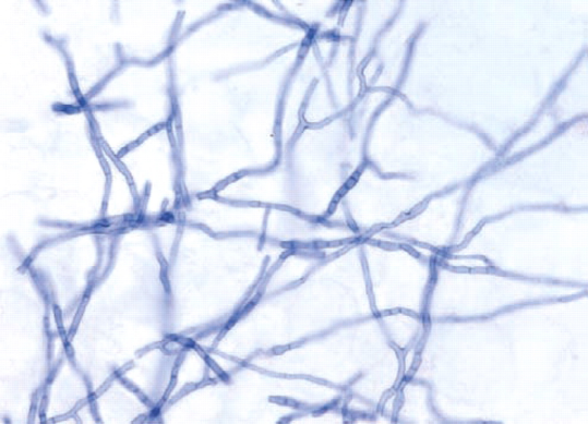
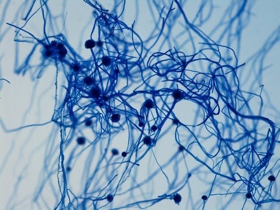
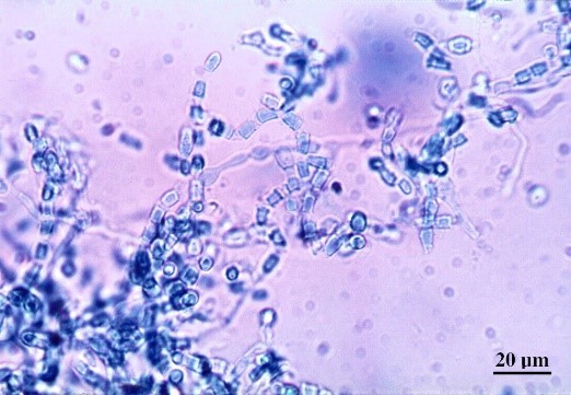
Moulds
Moulds grow as branching filaments called hyphae. These are long, thin cells which efficiently help the fungus to invade tissue. A mass of hyphae bunched together is termed a mycelium. Under favourable conditions (cooler than the human body and in the presence of air), moulds can also produce sporing structures. Spores (also called conidia in e.g. Aspergillus) are similar to seeds as they enable the fungus to reproduce and spread.
Endemic Dimorphic Fungi
Dimorphic fungi grow as moulds in the environment and as yeasts in human tissues. They are primary pathogens (can cause infection in immunocompetent individuals) and endemic to specific areas (mainly Americas).


Reach your personal and professional goals
Unlock access to hundreds of expert online courses and degrees from top universities and educators to gain accredited qualifications and professional CV-building certificates.
Join over 18 million learners to launch, switch or build upon your career, all at your own pace, across a wide range of topic areas.


