
Recording Teeth and Dental Pathology

Dental disease is routinely studied in both archaeological and modern skeletons because it is such a rich source of information. Not only can dental disease be useful in helping to identify an individual, but it can also be informative about diet, access to dental hygiene and care (or lack thereof), and even environmental conditions during the development of the teeth in childhood. As you will see next week, they are also an important source of biomolecular data.
Recording Dental Pathology
Before recording any signs of pathology, it is important to accurately identify any teeth that might be present. Teeth can be easily lost in the burial environment or during excavation and storage of remains due to their small size. This is especially the case for non-adults, whose teeth have not yet fully formed. Usually, an inventory of the teeth is performed along with the rest of the skeletal inventory. As with the rest of the skeleton, preservation and completeness of the teeth can have a significant impact on the anthropologist’s ability to analyse them. There are many methods and systems to record teeth and dentitions, but all include the following information:
- Tooth type (Incisor, Canine, Premolar, Molar)
- Tooth location (Maxilla or Mandible AND right or left side of the jaw)
- Tooth position (1st or 2nd incisor, 1st or 2nd premolar, 1st, 2nd or 3rd molar)
- Primary (deciduous/milk) tooth or Permanent tooth
It is important to correctly identify the individual teeth, especially if they have become removed or isolated from the jaw.
Dental caries (Cavities)
Most people have experienced a cavity, or dental caries, at some point in their lives. Caries occur when acid generated from bacteria in the mouth is able to break down and decay dental enamel and other tooth structures. Because sugar acts as a food source for the bacteria in the oral environment, high rates of caries have been associated with diets high in sugar or a lack of access to dental hygiene and care. Although caries formation takes time and goes through several steps, they are most often recognised as black-coloured holes in the teeth. These can range from very small to quite large. As previously mentioned, most people today have their dental caries filled by a dentist and these fillings can be useful for identification.
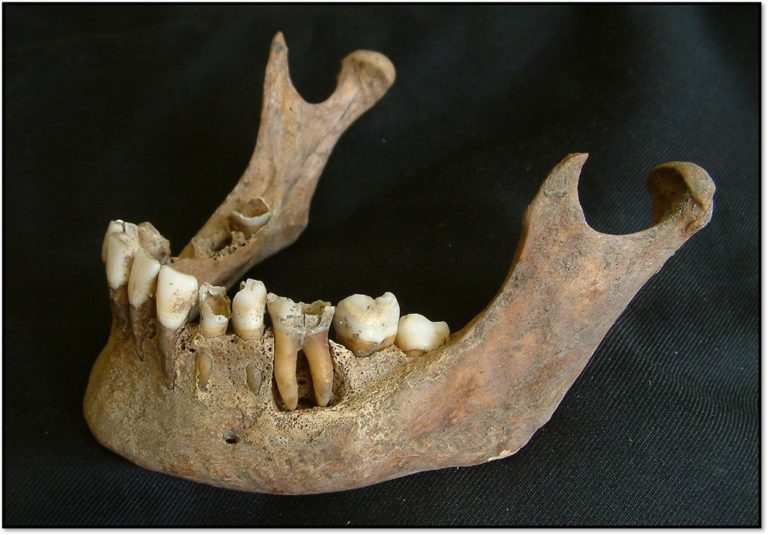
 A mandible showing several dental caries in the molars.
 A maxilla with severe dental caries in the molars.
If caries are left untreated they provide a way for infection to enter the jaw via the pulp cavity. This can lead to a periapical abscess, which appears in dry bone as a large rounded cavity beneath the tooth root. It can be extremely painful (see below).
 A periapical lesion, in this instance an abscess, evident from the remodelled edges of the lesion
Poor dental hygiene can of course ultimately lead to tooth loss. When a tooth is lost during life, the tooth socket will remodel and gradually close up (see below).
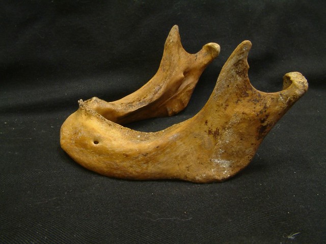 Extensive ante-mortem tooth loss in a lower jaw
Enamel Defects
Dental enamel is a special tissue in the body, as once it is formed, it does not remodel or heal like the rest of the body. Due to its high mineral content, dental enamel also preserves incredibly well, making it particularly useful in archaeological and forensic contexts. Defects in the enamel can be formed as a result of inadequate nutrition or severe illness during the development of the tooth in childhood. If this happens, the tooth continues to develop but lays down comparatively poor quality enamel, which results in the appearance of pits or lines in the enamel. If these enamel defects are present, it indicates that the individual endured some period of physiological stress (e.g. illness or malnutrition) during their childhood.
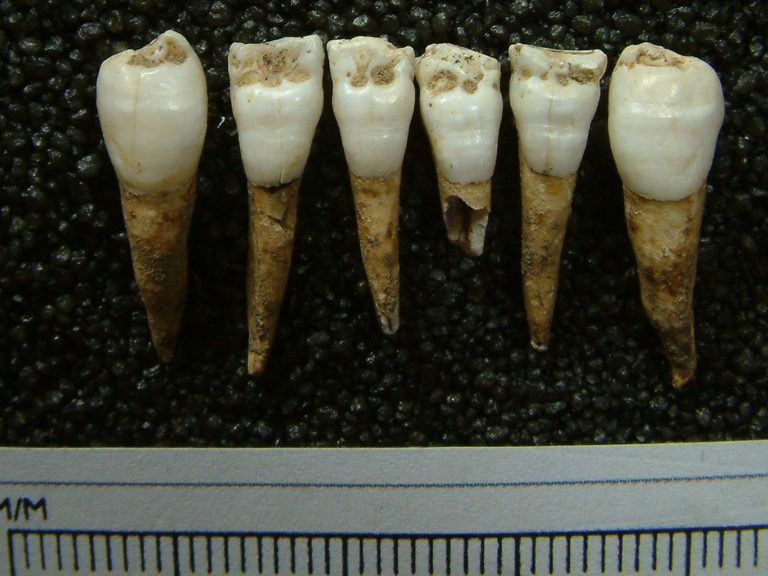
Dental defects in the enamel of the anterior teeth of a child.
Do you have any unusual or distinctive dental features that are unique to you? Have you received dental interventions in the past that might still be visible in your teeth or jaws today? If so, please add comments to the section below.
Practice identifying some dental disease using the 3D Models below in the ‘SEE ALSO’ section.

Reach your personal and professional goals
Unlock access to hundreds of expert online courses and degrees from top universities and educators to gain accredited qualifications and professional CV-building certificates.
Join over 18 million learners to launch, switch or build upon your career, all at your own pace, across a wide range of topic areas.


