
Skeletal Pathology in Forensic Investigation

Evidence of pathology and trauma on the skeleton provide important information that can help to identify the deceased and can provide possible evidence of the circumstances or cause of death. Comparison of skeletal pathology (e.g. a healed fracture to the arm) to ante-mortem medical records can provide important evidence for establishing individual identity.
Skeletal pathology can prove to be particularly important in forensic contexts in cases where other aspects of the osteological profile are very similar. For example, in war graves, most of the deceased are the same sex and similar age and are therefore not easily distinguished based on age and sex alone.
The identification and recording of pathological features on bones can be useful for the following reasons:
- Pathological conditions that affect for example facial features, or overall appearance, can help establish identity.
- Dental pathologies and treatment can affect a person’s appearance or be linked to ante-mortem dental records.
- Degenerative changes (e.g. joint disease) that can impact on an individual’s movement or lifestyle can be useful for identification.
- Other physical impairments (disabilities) which create individualising features (e.g. scoliosis) can be linked to medical records.
- Some features can hint at personal lifestyle factors, such as tobacco staining on teeth.
- Genetic diseases can be used to indicate familial links, and some may be associated with particular parts of the world – e.g. thalassaemia.
- Evidence of trauma can yield information regarding the cause of death and also post-mortem treatment of the body.
Bone structure and pathology
Before discussing abnormal bone, it is useful to consider the structure of normal bone. The outer layer of bone is called the periosteum. This can become inflamed in response to trauma or infection and produce a bony reaction – often referred to as periosteal new bone formation.
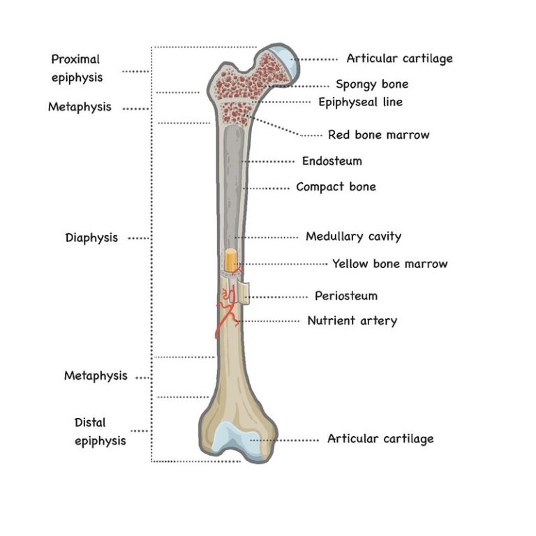
The periosteum covers a layer of cortical or ‘compact’ bone, which is dense and provides mechanical strength. The thickness of this compact bone varies with the skeletal element, depending on the function of different parts of the skeleton. For example, the weight-bearing bones will have more compact bone than other elements. If this compact bone becomes infected then it can produce a response referred to as osteitis. The bone usually appears thickened and denser in these areas.
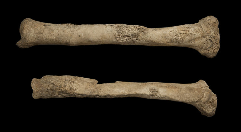 Right and left tibia showing a thickened appearance in response to infection
When the bone marrow becomes infected, either through an infection in the bloodstream or direct infection via an open fracture, infection may build up within the bone marrow, killing areas of bone around it and eventually draining out through a drainage channel (fistula). This is referred to as osteomyelitis. The bone will look very thickened and a cloaca (hole) may be visible on the outer surface where the infection has drained.
Bone Response to Pathology
Bone has a limited response to a wide variety of pathological conditions and this can complicate the diagnosis of a specific disease. Contrary to popular opinion, the human skeleton is not inert – bone is constantly remodelling (i.e. old bone is destroyed and new bone is formed). Pathological or traumatic processes affect the normal balance between osteoblast (bone forming cells) and osteoclast (bone destroying cells) activity. As Don Ortner (2003) discusses in his excellent book on skeletal pathology, this disruption in this balance can result in:
- Abnormal bone destruction
- Abnormal bone formation
- Combination of bone formation and destruction
- Abnormal bone size
- Normal bone quality but abnormal shape and contour
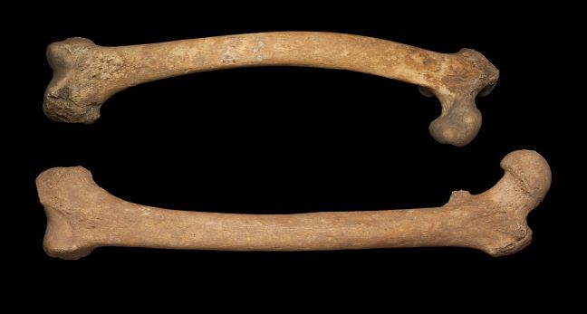 Femora from two different individuals. The femur on the bottom is normal and the femur on the top shows shape changes associated with vitamin D deficiency in childhood.
Take a look at the 3D model of a tibia with osteomyelitis below in the ‘SEE ALSO’ section.
A clear and comprehensive understanding of normal bone anatomy and human variation is critical to assessing pathology in the skeleton. So keep the concepts from this step in your mind as you move on to the next section, where you will learn more about the different types of bone response to pathological conditions.


Reach your personal and professional goals
Unlock access to hundreds of expert online courses and degrees from top universities and educators to gain accredited qualifications and professional CV-building certificates.
Join over 18 million learners to launch, switch or build upon your career, all at your own pace, across a wide range of topic areas.


