
Setting the scene: Pre treatment
Entering a radiation therapy department can be daunting. There are many different areas and units for you to attend. In the next two articles we will take you through a typical radiation therapy department so that you know what to expect when you arrive. This article brings you through a typical radiation therapy department in Ireland; however, this may differ from a department in your own country.
These photos were taken with the kind permission of St. Luke’s Radiation Oncology Network, at St. James’s Hospital campus in Dublin, Ireland.
Pre Treatment: Clinic room
Your first visit, before you start your treatment, will be in the clinic room where you will meet your radiation oncologist and radiation oncology nurse.
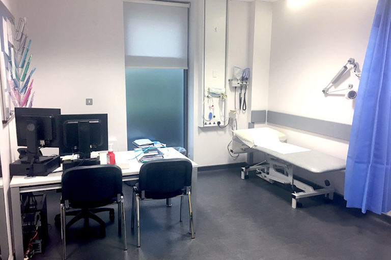 Clinic room
At this consultation, the radiation oncologist will discuss your treatment with you, the likely outcome, the anticipated side effects and how they will be managed. You will also have the opportunity to ask any of your questions. The radiation oncologist will seek consent for your treatment and may perform a physical examination.
The radiation oncology nurse will also take some details from you, especially about your past medical history, your family life and what supports you have to help you during your treatment.
Tips
- Bring the list of questions from earlier in the course with you to this consultation (as well as a pen and paper) to write down anything important you’d like to remember.
- Bringing a partner, relative or friend to the consultation can also be helpful in remembering the answers to your questions as well as to support you.
Pre-Treatment: Computed Tomography (CT)/Magnetic Resonance Imaging (MRI)
Once you have finished your consultation with the radiation oncologist, you will move to the pre-treatment area. Here you will be having a CT and/or MRI scan.
- An MRI scanner is used to take images of soft tissue (such as muscles and organs).
- A CT scanner, on the other hand, is useful for visualising bone and providing density information for planning radiation therapy treatments.
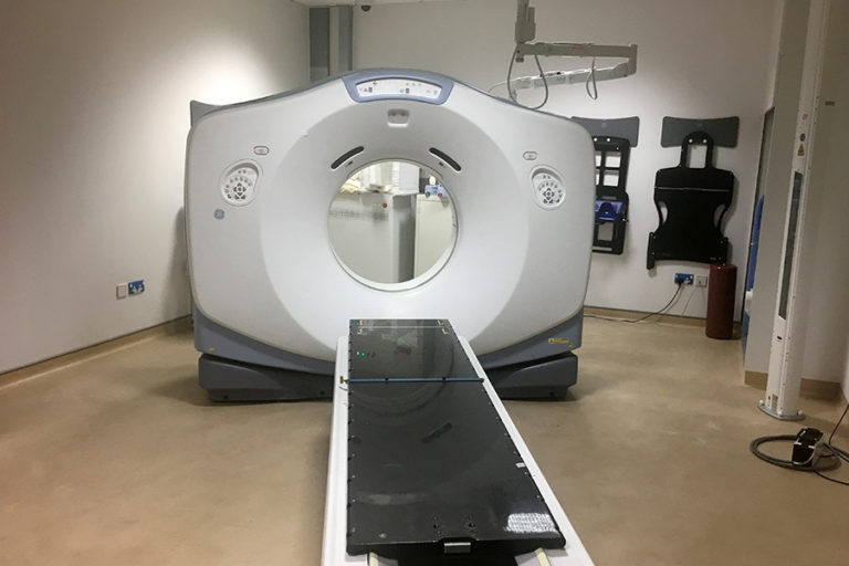 CT Scanner
Many patients wonder why they have to have further scans in the radiation therapy department when they have had previous scans. The scans in the radiation therapy department are taken with the patient in the treatment position and used to plan your specific treatment. This position is specifically selected to ensure that the linear accelerator can:
- Deliver the maximum dose to the tumour
- Minimise the dose to surrounding normal tissue
- Reduce side effects
Before the scan begins, you may be given an injection of dye into your arm called contrast. This highlights the lymphatic vessels and is very helpful to plan your treatment. If you are having a contrast injection, the radiation therapists will ask you some questions about your allergies. This is completely normal. Similarly, all women of child-bearing age will be asked if they may be pregnant prior to CT scanning. This is standard best practice.
In order to assist you to maintain this position, the radiation therapists in CT or MRI will use immobilisation devices. These can range from:
- Masks for head and neck treatments.
- Boards that tilt with cups for arms for breast, lung and oesophageal cancers (called breast boards or wing boards).
- Devices that are placed under the knees and feet for pelvic tumors, such as prostate, rectal and gynecological cancers (called knee or foot fixes).
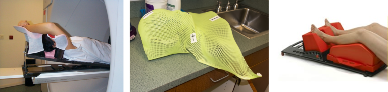 L to R: Immobilisation devices – Breast board, mask, knee/foot fix
The radiation therapists will spend quite some time selecting your position and immobilising you.
This is because the position selected here is very important as it will be the same position that you will lie in each day for every treatment.
The CT scan itself only takes about 10 minutes. If you have had an injection of contrast, it may feel cold in your arm as it is injected. Otherwise, you will feel nothing. The couch that you are lying on will move through the doughnut shaped dome of the CT scanner, but you will not be enclosed.
An MRI scanner can look similar to a CT scanner, however it is different and is used to image soft tissue (muscles and organs) within the body. The couch you are lying on moves through a short tunnel and can be enclosed. As MRI scanners can be quite noisy, you may be offered some ear protectors, such as ear plugs or headphones to wear during the scan. Some patients find MRI scanners difficult as many are enclosed.
Tips
- If you are claustrophobic, it is a good idea to inform the radiation therapists ahead of the scan.
- There are methods of helping you to relax and assisting you through this scan. Radiation therapists can speak to you via an intercom, or music be played to you through headphones (see note below).
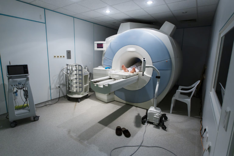 MRI Scanner (stock photo)
Following the scan, you will be free to leave the radiation therapy department as your pre-treatment process is complete.
Pre-treatment: Creating your personalised treatment plan
In the weeks between this scan and the start of your treatment, the radiation oncology team, that is, the radiation oncologist, medical physicist and radiation therapist, are producing a personalised plan for your treatment.
This involves delineating or drawing the tumour (known as the target), and all sensitive normal tissue (known as organs at risk) on your CT and/or MRI scan.
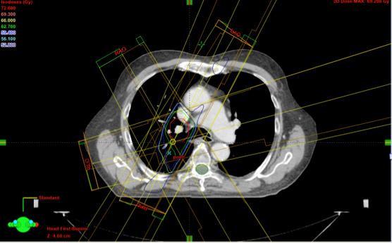 This is a treatment plan for a lung cancer patient. The tumour or target is delineated in red. The spinal cord, which is an organ at risk, is delineated in green.
Following this, beam angles and orientations are decided upon and doses calculated in a treatment planning system. Many independent checks have to be performed on your treatment plan before your treatment can begin. This is part of the routine quality assurance associated with radiation therapy.
In the next step, we will be looking at the locations for your radiation therapy treatment.
Please note that the wearing of headphones is not possible for those who have to wear masks (brain tumours or head and neck cancers) for their scans.
An Introduction to Radiation Oncology: From Diagnosis to Survivorship


An Introduction to Radiation Oncology: From Diagnosis to Survivorship
Reach your personal and professional goals
Unlock access to hundreds of expert online courses and degrees from top universities and educators to gain accredited qualifications and professional CV-building certificates.
Join over 18 million learners to launch, switch or build upon your career, all at your own pace, across a wide range of topic areas.


