
Array CGH: Identifying chromosome dosage abnormalities
As we discussed earlier in the course, chromosomes are essentially packages of DNA.
To revise, humans have 46 chromosomes or 23 pairs, with one chromosome of each pair inherited from each parent. Pairs 1-22 are the same (called the autosomes) whilst the 23rd pair (the sex chromosomes) comprise two X chromosomes in a female and an X and a Y chromosome in a male.
Diagnostic laboratories have increasingly been using array CGH (array comparative genomic hybridisation) as the first line investigation for patients with intellectual disability, developmental delay, autism and congenital abnormalities. It is also being applied in the prenatal setting following the detection of ultrasound scanning abnormalities in the pregnancy.
Array CGH has superseded the previous technique of examining the chromosomes down a microscope. This is primarily because it interrogates the chromosomes at higher resolution detecting loss (deletions) or gain (duplications) of all or part of a chromosome down to less than 100kb (kb = kilo bases. 1 kilo base = 1000 bases of DNA).
However, array CGH cannot detect chromosome rearrangements such as balanced translocations which still require microscopic analysis, and is unable to identify DNA sequence changes of an individual gene. Such changes are identified by a different method called gene sequencing.
How does array CGH work?
The patient and reference DNA are labelled with different coloured fluorescent dyes and applied to an array slide on to which is spotted DNA representing the whole genome. The patient and reference DNA binds to the DNA on the slide.
Gains and losses of patient DNA can be detected by identifying differences in fluorescence between patient and reference samples (see Figure 1). We have also included a PDF version of Figure 1 in the downloads section below.
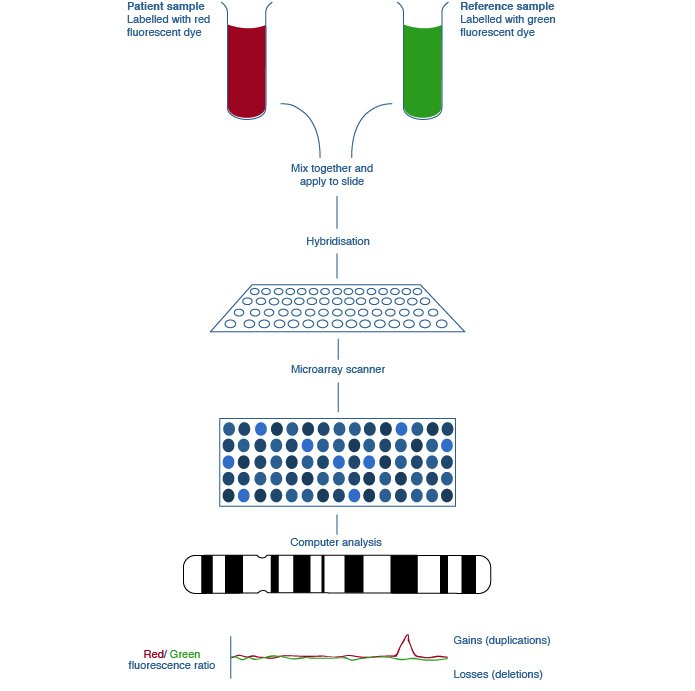
Figure 1: The array comparative genomic hybridisation (array CGH) process Click to expand
© St George’s, University of London
Talking point
What would you imagine are the challenges of Array CGH in clinical practice? What do you think it can and can’t be used for?
Please post your comments and we’ll consider these questions further in the next couple of steps.
The Genomics Era: the Future of Genetics in Medicine


The Genomics Era: the Future of Genetics in Medicine
Reach your personal and professional goals
Unlock access to hundreds of expert online courses and degrees from top universities and educators to gain accredited qualifications and professional CV-building certificates.
Join over 18 million learners to launch, switch or build upon your career, all at your own pace, across a wide range of topic areas.


