• The Open University

Histology: Using Microscopy to Study Anatomy and Identify Disease

Get an introduction to histology – the study of anatomy at a microscopic level – and use histopathology to identify disease.

23,770 enrolled on this course

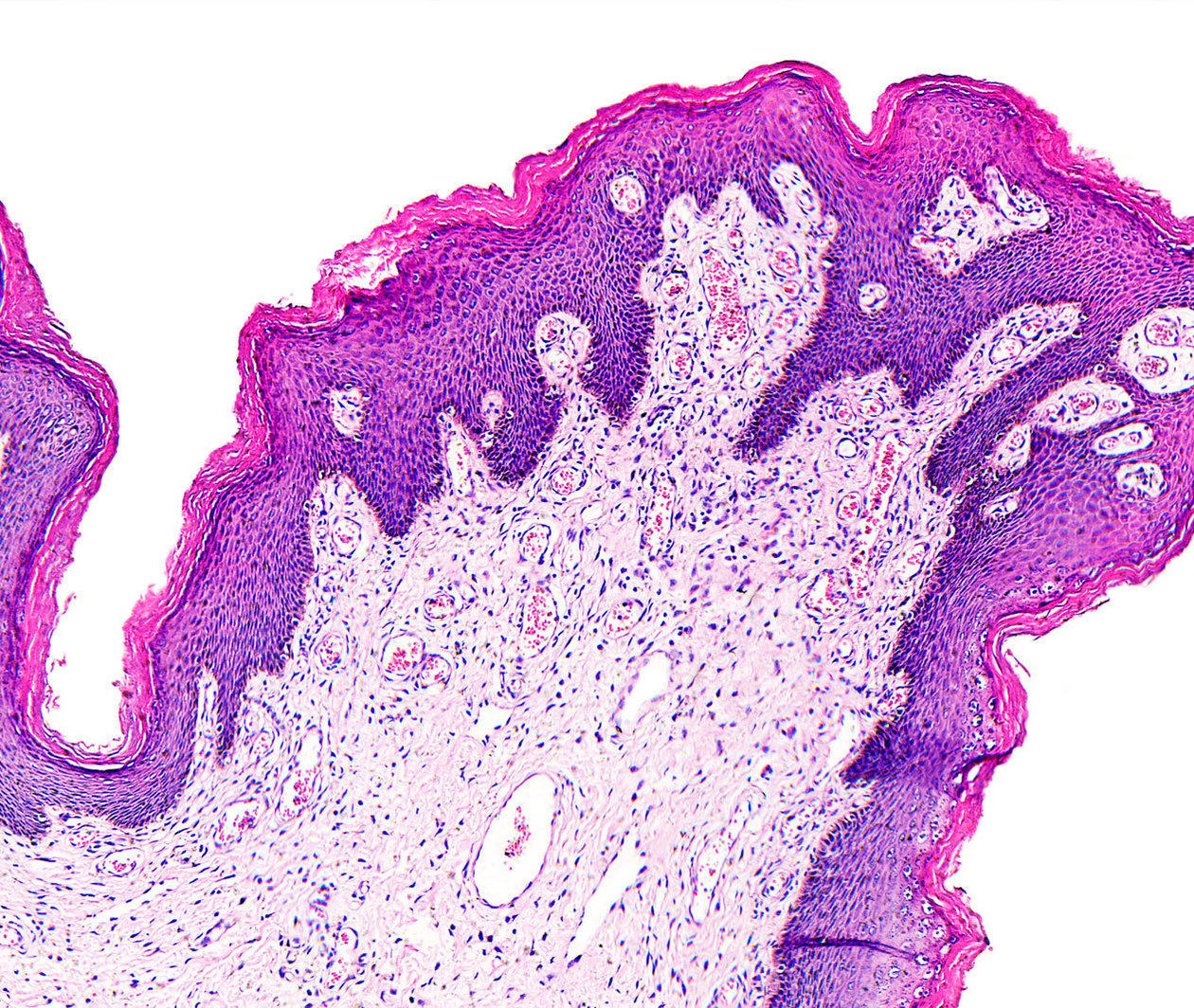
A microscopic view of tissue, as used in histology and histopathology

Histology: Using Microscopy to Study Anatomy and Identify Disease

23,770 enrolled on this course

  • 4 weeks

  • 3 hours per week

  • Digital certificate when eligible

  • Introductory level

Find out more about how to join this course

Examine tissues and identify disease with histology and histopathology

Histology is a key part of modern medicine – it helps us study cells and tissues at a microscopic level, and accurately diagnose diseases such as cancer.

This online course introduces the field. It starts with the basic principles of light microscopy, before focussing on the structure and function of human tissues, and the relationships between them.

You’ll use a virtual microscope to compare normal tissues with pathological (or diseased) ones, and use histopathology to identify and diagnose each disease.

The course was produced with the kind support of Dangoor Education.

Skip to 0 minutes and 14 seconds PROFESSOR: A career in histology and histopathology is rewarding, complex, and important. It’s used in biomedical research laboratories such as this one. It’s also used in hospital laboratories for the diagnosis of disease. But some of you may be wondering what histology and histopathology actually is. Well, it’s the study of tissues and diseases underneath the microscope.

Skip to 0 minutes and 41 seconds NARRATOR: This course will explain how to use a microscope, identify different tissues and some distinct pathological changes. The structure of a tissue is related to its function. Diseases produce characteristic changes in the appearance of a tissue. Much of the teaching is done through a virtual microscope, which gives you the feel and functions of the real thing. It draws on a large collection of sections collected from school, university, and hospital laboratories, so it’s your gateway to a huge collection of quality slides, making this course a unique introduction to histology.

Skip to 1 minute and 26 seconds PROFESSOR: Histology is taught in schools, both at GCSE and A-level. It’s also taught in biological science departments at universities, and it’s used in diagnostic laboratories in hospitals by people ranging from research technician right up to consultant histopathologist. This course will engage anyone who is interested in biomedical research or the diagnosis of disease. It will give you some insight into what it’s like to work in a research laboratory such as this one, or a hospital diagnostic laboratory.

Syllabus

  • Week 1

    Histology

    • Introducing histology

      Histology is the study of tissues and their structure and histopathology labs are found in most hospitals. See a histology department in action and discover how histological slides are produced.

    • Using a light microscope

      Histology departments use light microscopes with transmitted light. You’ll have the opportunity to put your learning into practice by using the virtual microscope tool to explore some real samples.

    • Measurements on the microscope

      Sometimes it is important to measure the size of a cell, the distance between cells or the number of cells in a given area. Have a go at measuring using the virtual microscope.

  • Week 2

    Tissues and cells

    • Identifying cells

      Cells in the body differentiate into many types as they develop, and within a single organ there may be many different types of cell. Learn how to recognise common cell types.

    • Identifying tissues

      Learn to identify different types of tissue using evidence provided by the virtual microscope.

  • Week 3

    Tissue structure and function

    • Relationship of tissue structure to function

      Look at a selection of tissues which have different functions and aim to relate the function to the structure of the tissue, seen under the microscope.

    • Secretion

      Examine two secretory tissues, the thyroid and the breast.

    • Movement

      Examine the function and structure of muscles needed for movement.

    • Structure and strength

      Tissues such as bone and cartilage give our bodies structure and strength.

    • Excretion

      The kidneys are excretory organs that perform three main functions to produce urine, filtration, reabsorption and secretion.

    • Communication

      The nervous system is divided into two main parts, the central nervous system (CNS) and the peripheral nervous system (PNS). They provide communication for the body.

    • Normal tissues

      Recognise normal tissues and compare them.

  • Week 4

    Recognising disease

    • Infection and inflammation

      Infection can affect any tissue of the body, producing cell damage and inflammatory reactions.

    • Cell death and degeneration

      Cell loss occurs in many tissues with age but the effects are particularly notable in tissues that have a limited capacity for regeneration, such as nerves in the CNS, the retina of the eye and the sensory cells of the inner ear.

    • Tumours

      Cell division is normally a highly regulated process. Tumours disrupt this regulation.

When would you like to start?

Start straight away and join a global classroom of learners. If the course hasn’t started yet you’ll see the future date listed below.

  • Available now

Learning on this course

On every step of the course you can meet other learners, share your ideas and join in with active discussions in the comments.

What will you achieve?

By the end of the course, you‘ll be able to...

  • Apply and understand the basic principles of microscopy
  • Identify a number of the more common tissues from their histological appearance
  • Assess and understand the relationship between the structure of some tissues seen under the microscope and their function
  • Identify gross histological abnormalities, such as neurodegeneration, inflammation and cancer

Who is the course for?

This course is designed for medical laboratory scientists, and students who are studying human biology at university or school.

What software or tools do you need?

You’ll use a virtual microscope that will work on all modern browsers on both desktops and tablets. But we recommend completing the course on a desktop, so that you can see the tissues more clearly, and have course articles, images and videos open in different windows.

Who will you learn with?

I have been Professor of Biology at the OU since 1999, with a particular interest in the use of multimedia for teaching biomedical sciences. We research on nanocarriers for drug delivery to the brain.

Who developed the course?

The Open University

As the UK’s largest university, The Open University (OU) supports thousands of students to achieve their goals and ambitions via supported distance learning, helping to fit learning around professional and personal life commitments.

  • Established

    1969
  • Location

    Milton Keynes, UK
  • World ranking

    Top 510Source: Times Higher Education World University Rankings 2020

Endorsers and supporters

supported by

Dangoor Education logo

Ways to learn

Subscribe & save

Buy this course

Start learning today

Choose the best way to learn for you!

$349.99 for one year

Automatically renews

$109/one-off payment

Free

Develop skills to further your careerFulfill your current learning needTry this course - with limits
Access to this courseticktick

Limited to 4 weeks

Access to 1,000+ coursestickcrosscross
Learn at your own paceticktickcross
Discuss your learning in commentstickticktick
Certificate when you're eligibleDigital onlyPrinted and digitalcross
Continue & Upgrade

Cancel for free anytime

Ways to learn

Choose the best way to learn for you!

Buy this course

$109/one-off payment

Fulfill your current learning need

  • Access to this course
  • Learn at your own pace
  • Discuss your learning in comments
  • Printed and digital certificate when you’re eligible

Subscribe & save

$349.99 for one year

Automatically renews

Develop skills to further your career

  • Access to this course
  • Access to 1,000+ courses
  • Learn at your own pace
  • Discuss your learning in comments
  • Digital certificate when you're eligible

Cancel for free anytime

Start learning today

Free

Try this course - with limits

  • Limited to 4 weeks

Find out more about certificates, Unlimited or buying a course (Upgrades)

Learning on FutureLearn

Your learning, your rules

  • Courses are split into weeks, activities, and steps to help you keep track of your learning
  • Learn through a mix of bite-sized videos, long- and short-form articles, audio, and practical activities
  • Stay motivated by using the Progress page to keep track of your step completion and assessment scores

Join a global classroom

  • Experience the power of social learning, and get inspired by an international network of learners
  • Share ideas with your peers and course educators on every step of the course
  • Join the conversation by reading, @ing, liking, bookmarking, and replying to comments from others

Map your progress

  • As you work through the course, use notifications and the Progress page to guide your learning
  • Whenever you’re ready, mark each step as complete, you’re in control
  • Complete 90% of course steps and all of the assessments to earn your certificate

Want to know more about learning on FutureLearn? Using FutureLearn

Get a taste of this course

Find out what this course is like by previewing some of the course steps before you join:

Join the conversation on social media

You can use the hashtag #FLHistology to talk about this course on social media.