Skip main navigation

New offer! Get 30% off your first 2 months of Unlimited Monthly. Start your subscription for just £35.99 £24.99. New subscribers only T&Cs apply

Find out more

Vision threatening diabetic eye disease

Describing vision threatening diabetic eye disease and the simplified international classification of DR and diabetic macular oedema.
10.1
Consuela: Diabetic eye disease describes a group of conditions which affect people with diabetes, many of which can threaten vision. There are 3 groups of conditions that we need to consider Firstly, there is the ocular conditions
22.6
which are actually associated with diabetes: such as cataract and diabetic retinopathy. Secondly, there is a group of conditions
30.9
for which diabetes is a known risk factor: such as glaucoma. And thirdly, there is a group of ocular conditions
39.4
where diabetes is a possible risk factor: such as vascular occlusions and corneal disease.
46.7
Consuela: People with diabetes may actively only seek an eye examination when they experience visual problems. It is essential to consider the possibility of diabetes in all patients with unexplained change in refractive error, complaints of diplopia, cataract or retinopathy.
62.8
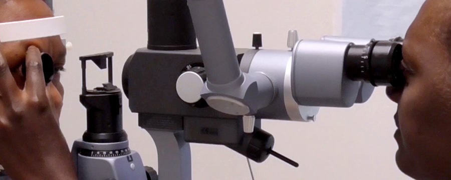
Charlotte: Gonna test your distance vision first
64.8
Consuela: Let us consider some of the likely presentations at the eye clinic. Fluctuating refractive errors Patients with newly diagnosed diabetes and unstable blood sugar levels can experience changes in refractive power of between 3 to 4 dioptres of far- or near sightedness. This is thought to be due to the effect of glucose products on the lens, resulting in temporary blurred vision. Reassuring the patient and educating them about good glycaemic control is essential before prescribing any additional spectacle correction.
99.6
Diplopia or double vision can arise from palsies of the cranial nerves. Diabetes is the cause of diplopia in 30% of people over 45 years and older who have developed acute extraocular muscle palsy. The underlying cause is thought to be microvascular damage to the cranial nerves. With good glucose control, people generally recover muscle function within 3 months.
126.6
The most important vision threatening diabetic eye diseases include cataract, glaucoma and diabetic retinopathy, including diabetic macular oedema. Other conditions, for example anterior ischaemic optic neuropathy, can affect a small percentage of people with diabetes.
145.9
Its essential that diabetes eye services closely monitor people with diabetes to identify vision threatening diabetic eye disease early on and prevent blindness. Cataract. Patients with diabetes are 2 to 5 times more at risk of early cataract formation. Its estimated that up to 20% of all cataract operations are on people with diabetes. Posterior subcapsular cataract is a major cause of vision impairment in people with diabetes. The risk of developing this condition increases with increasing duration of disease and severity of hyperglycaemia. The level of visual complaint should correspond to lens opacification. Comprehensive examination must always be undertaken to identify any underlying diabetic retinopathy in people with diabetes. Vision loss from cataract is reversible with surgery.
199
However, it is important to be aware that pre-existing diabetic retinopathy may worsen after cataract surgery.
207.2
When managing cataracts in people with diabetes, its also important to consider endophthalmitis, a severe infection within the eye and a devastating postoperative complication. People with diabetes are at increased risk of developing endophthalmitis after eye surgery.
224.8
Primary open angle glaucoma (POAG) involves a spectrum of disorders which are typified by characteristic changes on the optic nerve and visual field loss. A recent systematic review highlighted that people with diabetes have an increased risk of developing POAG. There is nearly double the risk of glaucoma in adults with diabetes.
246.8
Screening for glaucoma is challenging. However, it is important to carry out the detailed eye examinations essential for early detection of glaucoma as the disease remains asymptomatic until the late stages and often remains undetected by patients until fairly advanced.
264.3
Neovascular glaucoma is a visually serious consequence of underlying, systemic ocular disease. Inadequate blood supply leads to neovascularisation of the iris or neovascularisation of the lens for drainage in the eye and this ultimately leads to increased pressure. Diabetes accounts for approximately one-third of cases of neovascular glaucoma. It occurs particularly in those who have proliferative diabetic retinopathy. The incidence of neovascular glaucoma in diabetes is further increased amongst those who have already undergone previous ocular surgery. Panretinal photocoagulation is the procedure of choice for managing neovascular glaucoma. Diabetic retinopathy including diabetic macular oedema and proliferative disease is the most common complication of diabetes and the leading cause of visual loss.
313.8
Systemic blood pressure has a profound effect on the onset and progression of diabetic retinopathy and should be checked in every individual at each visit to the eye services. Over 90% of visual loss from diabetic retinopathy can be prevented with adequate treatment. People with diabetes may not be aware of retinal changes and therefore screening programmes are essential for early detection of diabetic retinopathy.
341.1
A workable classification system is essential to identify and characterise and grade the level of diabetic retinopathy found in screening programmes. Grading the changes seen in diabetic retinopathy screening describes the clinical severity of the condition and can be used to predict the risk of vision loss and provide appropriate management guidelines. Various diabetic retinopathy classification systems are in use worldwide. The system presented here is the International clinical disease severity scale for Diabetic Retinopathy and Diabetic Macular Oedema. This classification is simple to use, is primarily based on clinical examination and emphasises the severity of the clinical disease in the retina and specifically at the macula. The international clinical disease severity scale for diabetic retinopathy has five stages.
393.6
Standard colour fundus photographs of each stage are available to compare with the clinical findings. In Stage 1, No apparent retinopathy there are no diabetic fundus changes.
408.1
In Stage 2, which is Mild non-proliferative diabetic retinopathy, a few microaneurysms are present.
417.8
Stage 3 is called Moderate non-proliferative diabetic retinopathy. Here, we see the presence of microaneurysms, intra-retinal haemorrhages or venous beading which is more than just microaneurysms but less than severe non-proliferative diabetic retinopathy.
435.3
Stage 4, Severe non-proliferative diabetic retinopathy, is the most important stage to identify as treatment at this stage can prevent vision loss.
446
THE 4:2:1 rule is sometimes used to help identify Stage 4 disease. This is when any of the following signs are seen but there are no signs of proliferative retinopathy. 1. More than 20 intra-retinal haemorrhages in each of four quadrants. 2. Definite venous beading in two or more quadrants, and 3. Prominent Intraretinal Microvascular Abnormality in one or more quadrants.
476.3
Stage 5 is termed Proliferative diabetic retinopathy. Retinopathy in this stage has progressed to neovascular changes affecting the iris, angle, optic disc or retina with complications such as vitreous haemorrhage or retinal detachment. Laser treatment is needed urgently at this stage to preserve sight.
498.5
The international clinical disease severity scale for diabetic macular oedema The grader notes whether macular oedema is present or absent on clinical examination by noting the presence and distribution of exudates at the macula. If exudates are present, they can be further classified into mild, moderate or severe depending on the relationship of the exudates to the fovea as compared to standard colour fundus photographs.
526.7
The ocular conditions where diabetes is a possible risk factor are vascular occlusions and corneal disease. Retinal vein occlusion (RVO) is the first condition. The signs of RVO can mimic that seen in diabetic retinopathy. Therefore, when people with diabetes present with acute vision loss and signs of diabetic retinopathy in one eye, RVO should be considered. Patients may require laser treatment to address the complications of neovascular glaucoma and macular oedema. Retinal artery occlusion (RAO) is the second type of vascular occlusion. Retinal artery emboli and occlusion have been shown in some studies to be more common in people with diabetes although the evidence is not consistent. The prevalence of diabetes in patients with RAO has been reported to be as high as 20%.
575.8
It may be that diabetes is only one of a range of cardiovascular risk factors for this group of conditions.
584.7
Ocular surface diseases. A variety of corneal abnormalities are seen in people with type 2 diabetes, such as dry eye syndrome, corneal erosion, persistent epithelial defects and corneal ulcers. People with diabetes who wear contact lenses are at particular risk of developing epithelial damage. Treatment is generally with artificial tears, contact lens and topical lubricants.
612.5
In summary. Diabetes has an impact on vision in many ways and clinicians need to be aware of the range of presentations and suspect diabetes as an underlying cause. People with diabetes have not always shared the same favourable outcomes after cataract surgery as their nondiabetic counterparts, and appropriate surgical, laser and medical treatment has to be made available. Diabetic retinopathy requires early detection through a specific screening programme, and the use of the international classification system for grading and assessment of disease severity.

In this video Dr Consuela Moorman, consultant ophthalmologist with the Oxford University Hospital Trust, UK, considers the diabetes related changes in the eye which can lead to vision loss. She also introduces the International clinical disease severity scale for Diabetic Retinopathy and Diabetic Macular Oedema, which provides guidance to eye health professionals on when to intervene with treatment to reduce the risk of vision loss.

Managing eye complications needs the attention and understanding of both the person with diabetes and the eye health professional. As you watch the video and read the infographic below, consider the key educational message/s that need to be given to people with diabetes about their eyes and diabetes.

Infographic: How diabetes affects the eye

You can download and use this simplified diagram to explain how diabetes affects the eye to people with diabetes who are at risk of visual loss.

(Click to expand) or (Download as PDF)

  • MICROANEURYSM– small deep red dots in the retina (not haemorrhages) but as “out-pouches” from damaged or weakened capillary walls. They may or may not be leaking
  • HARD EXUDATE– distinct yellow white deposits of lipids that have leaked from damaged retinal capillaries. They can be seen as small deposits, larger plagues or in distinct circinate pattern around the macular
  • HAEMORRHAGES– intraretinal bleeding may be ‘dot’, ‘blot’ or ‘flame’ shaped depending on their depth within the retina
  • COTTON WOOL SPOTS– greyish white fluffy patches of discolouration in the nerve fiber layer, linked with focal hypoxia and swelling of nerve fibres
  • MACULAR OEDEMA – swelling at the macular caused by leakage and build of fluid into the retina, affecting vision
  • VEGF – Vascular Endothelial Growth Factor. Stimulates the growth of new blood vessels
  • NEOVASCULARISATION– abnormal and fragile new blood vessel growth on the retinal surface, which can bleed easily, affecting vision.

Diabetes leads to high blood sugars (hyperglycaemia) in the body. Hyperglycaemia triggers a wide range of biochemical changes within the body’s cells that can lead to:

  • Swelling and shrinking due to changes in water content within cells (osmotic changes)
  • Cell damage or even cell death due to imbalances between production of free radicals and antioxidants to limit damage (oxidative changes or oxidative stress)
  • An inflammatory response

All these wide ranging biochemical changes due to hyperglycaemia can lead to structural and functional changes throughout the body, including the eye.

In the anterior segment (front) of the eye, osmotic and inflammatory changes can cause corneal epithelial damage, changes in refraction and early development of cataracts.

In the posterior segment (back) of the eye, microvascular changes can lead to:

  • Increased leakage from damaged capillaries. The capillary cell wall can be weakened, which leads to development of microaneurysms, or increased leakage of lipids causing hard exudate deposits, break open and cause dot and blot haemorrhages. At the macula the leakage can be extensive causing swelling (macular oedema), directly affecting vision.

  • Blockage of the capillaries can be due to a number of factors such as thickening of the basement membrane in the wall of the capillary, increased “stickiness” of platelets, changes in red blood cells. Blockages to the flow of blood in the retina leads to a loss of oxygen (hypoxia), affecting the function of the nerve fiber layers and photoreceptors. We observe these areas of hypoxia in the retina as cotton wool spots. Tissues that are low in oxygen trigger the release of vasucular endothelial growth factors (VEGF) which in turn cause new (but abnormal) vessels to grow (neovascularisation) on the surface of the retina. These can break and leak easily causing a range of complications from haemorrhages, retinal detachments and visual loss.

This article is from the free online

Diabetic Eye Disease: Building Capacity To Prevent Blindness
Created by
FutureLearn - Learning For Life

Reach your personal and professional goals

Unlock access to hundreds of expert online courses and degrees from top universities and educators to gain accredited qualifications and professional CV-building certificates.

Join over 18 million learners to launch, switch or build upon your career, all at your own pace, across a wide range of topic areas.

Start Learning now