
What screening tests are available?
The ideal diabetic retinopathy (DR) screening test has sensitivity of at least 80%, a specificity of at least 95% and a technical failure rate of less than 5% of images being ungradeable. The gold standard photography method for the detection of DR, as defined by the Early Treatment Diabetic Retinopathy Study (ETDRS) group, is stereoscopic color fundus photography in 7 standard fields (30°). This method is also useful for identifying DME and subtle retinal neovascularisation. However, both patients and the photographers find the 7 fields method time-consuming, uncomfortable and labour intensive.
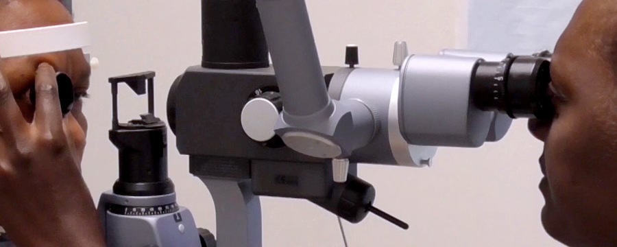
The International Council of Ophthalmologists’ diabetic eye care guidelines state, “Currently, the two most sensitive methods for detecting DR are retinal photography and slit-lamp biomicroscopy through dilated pupils. Both depend on interpretation by trained eye health professionals.” (ICO, 2017)
Professor Tunde Peto, clinical lead of Northern Ireland’s DR screening programme, introduces the video on this step which describes the test methods currently available to carry out DR screening. These features inform programmes’ decisions about which test is best suited for their specific setting. For example:
- Fundus photography creates a permanent record, and for that reason, it is the preferred method for retinopathy assessment. However, well-trained observers can identify DR without photography and there are many situations in which that will be the examination of choice.
- Using any instrument requires training and competence but more skill is needed to carry out indirect ophthalmoscopy and slit-lamp biomicroscopy than fundus photography. Newer, semi-automatic non-mydriatic fundus cameras can be easy to use.
- Media opacities in the eye – in the cornea, lens, or within the anterior or posterior chambers – will lead to photograph or view degradation and each one must be reviewed by trained personnel.
- The use of artificial intelligence to automate grading decisions is on the horizon, but this approach may remain out of reach for many health systems.
As you watch this video, consider what the most suitable DR screening method is for your setting. What are the main reasons for your choice?
Diabetic Eye Disease: Building Capacity To Prevent Blindness

Diabetic Eye Disease: Building Capacity To Prevent Blindness
Reach your personal and professional goals
Unlock access to hundreds of expert online courses and degrees from top universities and educators to gain accredited qualifications and professional CV-building certificates.
Join over 18 million learners to launch, switch or build upon your career, all at your own pace, across a wide range of topic areas.


