Skip main navigation

New offer! Get 30% off one whole year of Unlimited learning. Subscribe for just £249.99 £174.99. New subscribers only T&Cs apply

Find out more

Making treatment decisions

Dr Anthony Hall shares his perspectives and experiences as a vitreo-retinal surgeon in high and low resources health systems
10.9
Daksha Patel: So Anthony, as a clinician, when you have got a patient that’s been referred to you, say from a screening program, what are the key factors that you need to take into account when making decisions about the treatment?
29.1
Anthony Hall: There are multiple factors that would guide our decision on how to treat patients. These would include the setting that the patient is in– is it a high or low resource setting– and the stage of the disease. So if we look at the stage of the disease first, they may have severe pre-proliferative disease. They may have proliferative disease, or they may have diabetic macular oedema. Each of those stages will require a different approach. The resource setting will lead to the availability of different treatments. So in a high resource setting, you would have anti-VEGF drugs available, and these are often not available in a low resource setting.
69.2
Daksha: What would be the key initial assessment steps that need to be taken when making these treatment decisions for patients?
80.5
Anthony: There are two things we need to do at this stage. One is take a good history, and the second is to examine the patient. When taking the history, you’re looking at things that are going to modify the disease process, and this includes the general medical history. They may have a history of hypertension, and they may be pregnant. They may have renal failure. We’re looking at drugs that they may be taking, any anti-hypertensive drugs. Are they on lipidil, for example? Lipidil or fenofibrate has been shown in randomised controlled trials to reduce the need for treatment of diabetic maculopathy. And how is their glycaemic control? They may have a diary in the low resource setting.
120.7
They may even have access to a haemoglobin A1c, which will give us an idea of how well their control has been. When it comes to examining the patient, very critical is the visual acuity. Patients may come through screening with absolutely normal acuity. That’s the whole point of screening. We need to find them early. But if their vision is poor, there may be vitreous haemorrhage. There may be macular oedema, which will modify the way we’re going to treat them. The slit lamp examination of the interior segment is important. There may be a cataract interfering with their vision. They may have rubeosis because they have so much ischaemia.
155.3
We then move on to looking at the fundus and looking for evidence of that diabetic eye disease that has been referred to us. It may be maculopathy with exudates and haemorrhages. There may be severe non-proliferative disease or proliferative retinopathy. In high resource setting, we then have more ancillary investigations available to us. The most frequently used these days is optical coherence tomography, or OCT.
181.9
Fluorescein angiography is also important, although not everyone uses it. But there’s increasing evidence that fluorescein angiography will delineate areas of ischaemia both in the macular and peripherally. And the peripheral ischaemia is often driving the production of vascular endothelial growth factor, and that causes more macular oedema. So our understanding of macular oedema is increasing as we use more wide field angiography and understand the peripheral ischaemia. And the OCT will show the oedema very well and is absolutely critical in then following the patients in their treatment of that oedema. And many high resource settings will now have OCT angiography. This is quite new, and we’re learning more and more about how that will guide us in our treatment of these patients.
228.4
Daksha: In a low resource setting, how would the lack of equipment affect the manner in which you can take decisions to treat?
239.4
Anthony: We’re talking specifically about macular oedema here. The OCT makes it so easy to assess macular oedema. Clinically, it’s more difficult when you’re using a non-contact lens to image the fundus. So I encourage people to use a contact lens which makes it much easier to visualize the macular oedema. In practice, there are also a lot of other clues to the fact that the patient has significant macular oedema. You can see exudates encroaching on the fovea or even under the fovea. There are hemorrhages. There are micro aneurysms. These all indicate that they may be significant macular oedema present, particularly in the presence of low visual acuity.
279.3
I think in the low resource setting it would also be important to try and get some sort of record of the fundus using some of the fundus imaging systems that are now available for smartphones. And with that, you can then educate and counsel your patient as to the type of macular problem they have and watch them getting better. It’s very rewarding for the clinician and the patient to see those circinate exudates disappearing over time after some focal laser.
307.8
Daksha: In a clinical setting, you’re likely to be seeing a range of different stages for diabetic retinopathy. How do you go about making treatment decisions for each of those stages?
322.3
Anthony: The decision becomes more difficult when the patient has severe non-proliferative disease. And the decision will be based on the setting that the patient is in and personal factors that are affecting that patient. So the high resource setting, most patients would be inclined to come back. But you may find patients in a high resource setting who you can tell are going to have poor compliance because of perhaps their level of education, financial difficulties they may be having, and so on. And these certainly apply more frequently in the low resource setting. So if you feel that a patient may not return for their treatment or their review, then early treatment would be recommended.
367
On a personal disease level, patients may have pregnancy, may be pregnant, and they would certainly require very close following and monitoring and probably would benefit from early treatment in that situation. In macular disease, if a patient requires cataract surgery, you really need to get the macular disease under good control, because cataract surgery will certainly make the oedema worse. With proliferative or pre-proliferative disease and patients in the setting where you really are concerned about the compliance, I would be inclined to treat them early. Once the patients develop proliferative disease, then the decision is quite easy. And patients require treatment. In most settings, people would still recommend laser.
417.3
There is, however, some good evidence coming out that over a two-year period, anti-VEGF drugs are as effective as laser and perhaps produce better results in that they don’t affect the visual field. And visual outcomes appear to be better. The concern is that patients do need to keep coming back. So if there’s any concern about compliance, laser is still a very reliable treatment.
442.9
Daksha: On a practical level, what is the best approach for the treatment of diabetic macular oedema?
449.3
Anthony: This is going to depend, once again, on the setting and on the level of visual acuity. So in patients in a low resource setting, if they have a circinate exudate that’s threatening the fovea, then the treatment is effective and quite easy to apply. Treat the center of the circinate with the laser. If they have central macular oedema, then treatment with an anti-VEGF would be ideal. The grid laser treatments are definitely inferior to anti-VEGF drugs. But not all low resource settings are going to have anti-VEGF drugs available. So those patients would need grid treatment, recognizing that they’re not going to do as well as those patients who have access to anti-VEGF drugs.
499.6
In a high resource setting, the anti-VEGF drugs are the main way of treating patients. I believe Avastin has been added to the WHO Central Drug List. And this really is critical for our management of diabetic macular oedema. If we’re going to improve vision and prevent severe visual loss, it really is the way forward. And it’s much cheaper than the other anti-VEGF drugs that are available. The problems are being able to compound it correctly so that’s it in a sterile syringe. And if there are no compounding pharmacies available, then you might consider withdrawing multiple doses from the vial. And with a careful technique, this has been shown to avoid contamination of the vial and thereby prevent endophthalmitis.
553.2
So I really do believe that we need to make Avastin widely available in low resource settings so that all patients can get the treatment they deserve.
564.2
Daksha: In the decision to treat patients with diabetic retinopathy, what is the role of patient education in this process?
574.9
Anthony: It’s absolutely critical for every person living with diabetes to have a good understanding of how diabetes can affect their eyes. As an ophthalmologist, you don’t have a lot of time to explain how important screening is, to go into the disease process that affects the back of their eye, and to explain the treatment. And so a diabetic educator or a counselor who’s been trained to help patients or people living with diabetes to understand the disease process and to enable them to take control and manage it themselves is critical in this situation. They can explain the disease process. They can explain the treatment.
621
And it might take you a few minutes to give a very basic outline of what you’re going to do, but a special person in the clinic could spend a good half an hour really enabling a patient to understand and thereby take better control of their disease and be therefore more likely to comply with the treatment and comply with follow up.
642.9
Daksha: So finally, Anthony, you might be faced with a very difficult situation when you get to a point where you are unable to treat a patient. How do you go about this?
655.3
Anthony: This is always a difficult situation for the people looking after a person living with diabetes when they’ve got to that stage that they’re blind in both eyes. And I’d like to break it down, perhaps into a situation where the patient has lost vision in one eye and the decision on whether to treat them or not, and they’ll want to do the patient who has lost vision in both eyes and can’t be treated. So once again, it depends on the setting. Patients in a low resource setting who have a vitreous hemorrhage or a traction or detachment of the macula may not have access to the treatment.
690.1
And the vitreous hemorrhage may clear, but the traction or detachment of the macula is not going to get better on its own and ideally requires a vitrectomy, which is available in a high resource setting. And if that detachment is not a very old one, the outcomes can be quite good. However, long standing detachments wouldn’t be treated even in a high resource setting. We shouldn’t give up at that stage. There are rehabilitative services that can help patients in a high resource setting. They can be given incredible computer assistance with how to read, how to make use of their limited vision, mobility training. And these should be available in a low resource setting as well, to a certain extent.
729
Certainly the mobility training and the low visual aids are things we shouldn’t be forgetting about.
733.9
Daksha: Thank you very much.

To be effective, the strategies we use to detect and treat diabetic retinopathy must include the perspectives and needs of people with diabetes and align with what is practical and available in the local health system.

Mr Smith, is a 47 year old living with type 1 diabetes since the age of 17 years. He was referred from retinal screening at the age of 30 with exudates in the macula of his right eye. No immediate treatment was required but he was followed up regularly. The leak on the right macula increased 4 years after first presentation and was treated with focal (localised to the affected area) laser. Ten years later, his retinopathy had progressed with neovascularisation around both optic discs and a sudden haemorrhage in the retina.
Pan-retinal (all around the retina) photocoagulation laser treatment was started immediately, and despite the application of 3400 burns over 4 sessions in the left eye there was there was further areas of ischaemia and neovascularisation in the left retina. A similar amount of laser treatment was provided in the right eye. Both eyes also received two further laser sessions but both continued to show signs of activity with episodes of vitreous haemorrhage. Vitrectomy with endolaser (directly in the eye during surgery) was then performed in each eye. This resulted in stability to each eye with visual acuity of 6/9 in each eye.
Case study taken from “A practical manual of diabetic retinopathy management” edited by Peter Scanlon, Ahmed Sallam and Peter Van Wijngaarden

The treatment journey for many patients will be long. It may also be unsatisfactory, even in highly resourced health systems. Decisions on treatment need to take into account available resources and equipment, skills of the eye health team and the barriers faced by the patient in adhering to the treatment pathway.

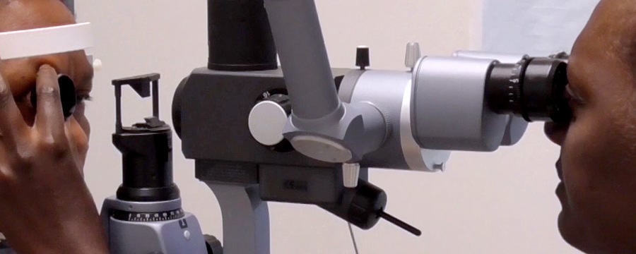
In this video, Dr Anthony Hall, consultant ophthalmologist, shares his perspectives and experiences as a vitreo-retinal surgeon in high and low resources health systems. As you watch the video, consider the local treatment challenges faced by providers and people with diabetes in your local setting.

This article is from the free online

Diabetic Eye Disease: Building Capacity To Prevent Blindness

Created by
FutureLearn - Learning For Life

Reach your personal and professional goals

Unlock access to hundreds of expert online courses and degrees from top universities and educators to gain accredited qualifications and professional CV-building certificates.

Join over 18 million learners to launch, switch or build upon your career, all at your own pace, across a wide range of topic areas.

Start Learning now