Skip main navigation

Different treatment options for diabetic eye disease

The range of laser treatments available to treat diabetic retinopathy and diabetic macular oedema and the use of anti-VEGF to treat proliferative DR
11.2
Once a patient has been identified as needing treatment for diabetic eye disease, the first step is to examine their retinas in detail using an indirect opthermascope and slit lamp biomicroscopy a Indirect ophthalmoscopy is useful for looking for evidence of peripheral neovascularisation, retinal detachment, and ischaemia (areas with capillary blockages). The macular is examined with the slit lamp and a variety of lenses. Non-contact indirect lenses are the most versatile and well-tolerated by patients. However, a contact lens will give a much better assessment of retinal thickening and oedema. Further useful investigations to guide the treatment process include colour retinal photographs, fluorescein angiography, and optical coherence tomography, OCT.
58.6
Color retinal photographs taken at screening are useful for canceling patients and following up the effects of treatment, for example, from lasers. They are not that good for monitoring diffuse macular oedema. Modern wide-field retinal imaging can identify more peripheral neovascularisation. Fluorescein angiography, or FA, is a useful diagnostic procedure which gives a real time assessment of the capillary blood flow in the retina. It precisely indicates the areas of leakage or blockages in blood vessels and capillaries. FA can help guide the pan-retinal photocoagulation laser when treating proliferative diabetic retinopathy. Peripheral ischaemia may have a role in driving macular oedema and ultra wide-field FA has been found to improve detection of peripheral ischaemia by 3.9 times.
105.8
Finally, in diabetic macular oedema, FA can identify areas of blockages in the capillary blood flow that may help explain visual loss and areas of leakage that would benefit from focal laser. OCT imaging provides a unique cross-section of the retina, showing how it’s structured. In a well-resourced eye clinic, most clinicians rely heavily on OCT scans to diagnose diabetic macular oedema, decide if treatment is necessary, plan treatment intervals, and assess if the treatment is working. OCT signs at presentation may even be used to give some idea of the likely visual prognosis. OCTs can also be shown to patients to explain the disease and share improvements. This helps with treatment compliance, as patients can see that they are slowly getting better.
152.6
Treatment options vary according to whether the patient has been identified with proliferative disease or maculopathy. Laser has a well-established role in treating diabetic eye disease. It’s particularly effective in preventing blindness in proliferative diabetic retinopathy using PRP. Over 3,000 laser burns are made to cover the peripheral retina, seal the leaking blood vessels, and stop new blood vessels from growing. Laser can be used to treat maculopathy, but this has largely been replaced in high resource settings by anti-VEGF drugs which give superior visual outcomes. Laser has poor outcomes for treating diffuse central macular oedema. This makes it a difficult treatment for patients to accept and doctors to advocate for.
194.5
It’s important to be aware of and inform patients of possible complications of laser treatment, including impairment of contrast sensitivity, color vision, and night vision. Occasionally, laser scars close to the macula can enlarge and involve the fovea and cause loss of vision. There are some specific issues in providing laser treatment in low and middle income countries. On the positive side, laser treatment means fewer treatment visits are required than for injections. In selected cases, it can be possible to stabilize the situation with one treatment. This is important for patients in remote settings with limited access to eye services. Excellent outcomes are possible in early maculopathy. However, on the negative side, purchasing a laser is expensive.
239.3
And there are many countries with no lasers. With the projected rise in the numbers of people needing treatment for diabetic retinopathy, there is an urgent need for health systems to invest in lasers and provide suitable training for ophthalmologists. Laser treatment is often poor quality when not provided correctly. The commonest mistake is not giving enough laser to remove the risk of progression of retinopathy. Laser is very time consuming and clinicians may have difficulty finding time to complete a number of PRPs in a day packed with other responsibilities. Improving a laser service. Laser is sight giving when given promptly and in sufficient quantity to treat proliferative disease. Prompt and fast treatment for patients can achieve improvement within a week or two.
283.9
It’s important to keep treating until new vessels have all resolved. Consider treating those patients who are unlikely to return in a single session. This may lead to some short term effects, such as making macular oedema worse. But it’s better than a patient blinded by inadequately treated proliferative retinopathy. Laser PRP is very effective and reduces severe visual loss to 4%. However, it may be ineffective in some eyes. And 5% of eyes need vitrectomy in spite of laser. The introduction of anti-VEGF therapy has revolutionized the management of diabetic macular edema. There are several anti-VEGF drugs in use at the present. Lucentis and Eyelea are intended purely for intraocular injection. And each vial can only be used for one patient.
328.8
Avastin was intended to be given intravenously as an anti-cancer drug, and it comes in vials of 100 milligrammes. The vial can be subdivided to treat up to 40 patients. As a result, a single dose of Avastin can work out to be over 100 times cheaper than a single dose of Lucentis. Indications for anti-VEGF drugs to treat diabetic eye disease. - Clinically significant macular oedema due to diabetic retinopathy. - Macular oedema, secondary to brunch or central retinal vein occlusion. - Vitreous hemorrhage, secondary to proliferative diabetic retinopathy. - Prior to vitrectomy for proliferative diabetic retinopathy. Not more than a week before surgery. Anti VEGF reduces the risk of intraoperative hemorrhage and retinal tears, and reduces usage of silicone oil. - Neovascular glaucoma.
377.6
Use of anti-VEGF at the time of cataract surgery may minimize the worsening of preexisting macular oedema. Several trials have compared anti-VEGF with laser treatment for diabetic macular oedema. People receiving anti-VEGF were approximately four times more likely to gain 15 or more letters of visual acuity at one year compared to people who receive laser treatment. They were also 90% less likely to lose 15 or more letters of visual acuity in one year. Anti-VEGF treatment is much more effective than laser for treating diabetic macular oedema. The challenge is to make it more universally available in low and middle income countries. Laser remains the gold standard for treatment proliferative diabetic retinopathy.
420.8
This is because PRP offers a permanent cure in one or two treatments, whereas anti-VEGF injections are ongoing. Even in high income countries, compliance with anti-VEGF injections is an issue. Patients may have one or two injections and then not return until they have suffered irreversible vision loss because the proliferative disease has progressed. There are considerable cost implications for health services and patients in adopting anti-VEGF. Patients need 10 to 14 injections over two years. The risk of endophthalmitis from injections is another very important consideration. When to use anti-VEGF drugs in low resource settings. As part of any established diabetic retinopathy screening and treatment programme where there will be enough patient demand to make it worthwhile.
466.9
In urban settings, with regular eye services and better follow up. In patients with diabetic macular oedema, who have already demonstrated good follow up or who have failed laser treatment. Anti-VEGF is not yet recommended for treating proliferative disease in low resource settings, due to poor compliance and the cost to the health service and patient. Avastin can be an option by getting compounding pharmacies to prepare individual doses. However, this does carry some risk. Outbreaks of endophthalmitis have been linked to pharmacy failures. There are alternatives to using a compounding pharmacy. Exact portions can be withdrawn without a compounding pharmacy. This carries a high risk of contamination. Pool patients over one day using multiple punctures from a vial.
512.7
This stretches human resources and often means no patient choice. Make multiple punctures from one vial until it is empty over a period of up to a week. There are obvious concerns about the safety of this technique. However, some recent studies suggest that the risk of contamination is very low if the file is correctly swabbed each time. In summary, Treatment of diabetic retinopathy starts with a detailed examination and investigation. Fluorescein angiography and OCT provide a detailed understanding of the pathology in the retina. The gold standard for proliferative diabetic retinopathy remains pan retinal photocoagulation. Laser treatment is preferred in low resource settings and when compliance for follow up is likely to be a problem.
556.4
Anti-VEGF is very effective for treating macular oedema, but the cost of treatment remains a challenge in low resource settings.
“In our clinic, a typical patient, Mrs X, was first seen with a visual acuity of 6/9, a few macular exudates, and proliferative disease. The treatment plan followed the textbook recommendation of doing focal laser for the maculopathy first. The patient then missed two appointments and pan-retinal photocoagulation (PRP) was delayed by about two months. When PRP was finally given, the intention was to give it in the recommended multiple sessions. However, due to further missed appointments, the interval between laser sessions was over a month. This allowed fibrovascular proliferation to continue. It was six months from the time of presentation before laser was completed. By then, tractional retinal detachment involving the macula had developed and vitrectomy was required. Mrs X’s final visual acuity was counting fingers at three metres.”

There are clearly established treatment plans in theory but in practice they require working closely with the patient.

Many patients are very anxious about having laser treatment and an adequate explanation of the process is essential. Most patients characterise the “pain” of laser treatment as brief, intermittent, sharp or piercing. Pain is also often described as being worse during re-treatment. It is important to manage patient’s anxiety throughout treatment and encourage them with the benefit of preventing long term vision loss.

Anti VEGF may not a suitable option for all patients or all settings.

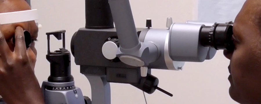
As you watch this video, consider what can be practically done to ensure patient adherence to treatment plans?

This article is from the free online

Diabetic Eye Disease: Building Capacity To Prevent Blindness

Created by
FutureLearn - Learning For Life

Reach your personal and professional goals

Unlock access to hundreds of expert online courses and degrees from top universities and educators to gain accredited qualifications and professional CV-building certificates.

Join over 18 million learners to launch, switch or build upon your career, all at your own pace, across a wide range of topic areas.

Start Learning now