
Estimating sex from the pelvis

The pelvis is one of the most sexually dimorphic regions of the skeleton. The overall shape of the male pelvis is narrow and steep, and the female pelvis is generally broader, with a larger pelvic outlet to facilitate pregnancy and childbirth. Since this is the only region of the skeleton where there is such a key functional difference, the pelvis proves to be one of the most important bone for determining biological sex. The accuracy of sex determinations from the skeleton based on the pelvis alone is usually placed at around 96%, but this will vary between populations as well as the degree of preservation and fragmentation (see this article by Inskip et al. 2019 for a recent summary and test of some of the methods). In forensic and archaeological contexts the pelvic girdle is often not complete and sex determination relies upon recording and assessing individual sexually dimorphic features as outlined below. The following criteria are described in most text books on skeletal analysis and here we have synthesised information from both the European and Anglo-American schools. A reference list is provided at the end.
If you’re unfamiliar with pelvic morphology, take a look at the annotated 3D models of male and female pelves first. These are located in the ‘SEE ALSO’ section below this article.
Sciatic Notch
The male sciatic notch tends to be narrow and ‘U-shaped’ and the female sciatic notch is wider and ‘V-shaped’. In some individuals, the sciatic notch may seem intermediate and difficult to assign as male or female. In such cases, it is useful to refer to the ‘composite arc’.
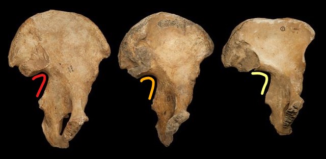 Three pelves with the sciatic notch highlighted. Male is on the left in red, intermediate in the middle in orange and female on the right in yellow.
Composite Arch
If you do have an intermediate sciatic notch, and you’re not sure whether it’s male or female, it’s useful to use the composite arch which is described in Bruzek 2002. Here, you follow the edge of the sciatic notch around the superior surface of the auricular surface. In males, you’ll see that it forms a continuous arch. When you try to do this in females, you’ll see that it misses the superior surface of the auricular surface, and it forms almost two separate arcs.
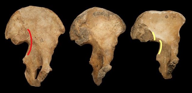 The composite arc highlighted with the single ‘male’ arc on the left and the ‘female’ double arch on the right. (Extra: Try to find the composite arch on the middle pelvis. Do you think it is male or female?)
Preauricular Sulcus
This is a depression in the bone surface located inferior to the auricular surface. Both males and females may have a preauricular sulcus, but it is less common in males. The morphology of the sulcus also differs between males and females. In females, it tends to be deeper and to have clearly demarcated edges, whereas in males it is usually shallow with open sides. More information is also provided in the Bruzek (2002) article cited above.
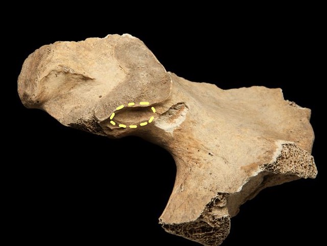 In this female pelvis, the preauricular sulcus is highlighted in yellow. Be sure not to confuse the preauricular sulcus with the nearby post-mortem damage (white-coloured depression).
Pubic Bone
Overall, the features of the female pubic bone tend to be more gracile (light, fine, smooth) and more sharply defined than in males. Male pubic bones are usually thick and more robust.
Subpubic angle
The angle formed by the ischio-pubic ramus below the pubic symphysis is wider and more ‘U-shaped’ in females than in males. Males have a narrower subpubic angle that tends to be more ‘V-shaped’.
 A female subpubic angle is highlighted in yellow and a male subpubic angle is highlighted in red.
Ventral Arc
Females have a ‘ventral arc’, which is a flattened, roughly triangular-shaped aspect to the ventral (anterior) surface of the pubic symphysis.
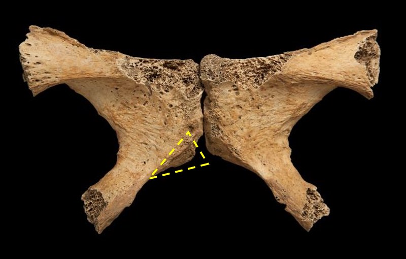 Female pubic bones with the ventral arc highlighted on one side.
Iliopubic ramus
In females, the iliopubic ramus (part of bone connecting the ilium to the pubic bone) is long relative to the diameter of the acetabulum. In males, the length of the iliopubic ramus and diameter of the acetabulum are often of roughly equivalent dimensions.
 A female and male pelvis highlighting the relative length of the pubic bone to the diameter of the acetabulum.
Ilium
In males, the ilium is narrower and the iliac blade has a more pronounced ‘S-shaped’. In females, the ilium is broader and the ‘S-shaped’ appearance of the iliac crest is shallow.
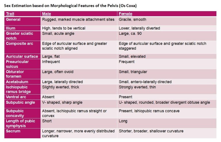
For a brief summary of these and some additional sexually dimorphic features of the pelvis, please see the table provided below.
Practice recognising and recording the traits used to estimate sex from the pelvis using the 3D models of the ox coxae in the SEE ALSO section below.
In the next step, you’ll be learning how to estimate sex from the skull, which has a different range of features, advantages and limitations.
References
Some of the general references below also include information about sex estimation from the skull.
This website provides a useful summary of methods http://talus.matrix.msu.edu/.
Buikstra, J., & Ubelaker, D. H. (1994). Standards for data collection from human remains (p. 44). Fayetteville, AR: Arkansas Archeological Survey Research Series
Ferembach, D., Schwidetzky, I., & Stloukal, M. (1980). Recommendations for age and sex diagnoses of skeletons. Journal of Human Evolution, 9, 517– 549.
Phenice, T. W. (1969). A newly developed visual method of sexing the os pubis. American Journal of Physical Anthropology, 30, 297– 302.
White, T., Black, M., Folkens P. 3rd Edition (2005) Human Osteology. Elsevier.


Reach your personal and professional goals
Unlock access to hundreds of expert online courses and degrees from top universities and educators to gain accredited qualifications and professional CV-building certificates.
Join over 18 million learners to launch, switch or build upon your career, all at your own pace, across a wide range of topic areas.


