New Forensic Research on Burned Bone

Examining heat-induced change in bone
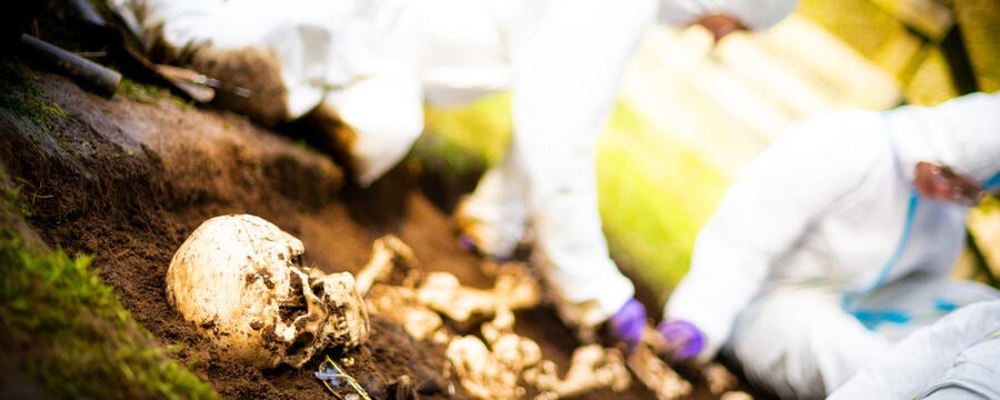
Over the past 10 years or so, more and more researchers have been analysing burned bone. We’ve realised that despite the significant problems that such heat-induced changes introduce to the identification process, the changes themselves can be useful to investigate. We now have a range of new methods for exploring these changes, many of which focus on the microstructure of bone. This is really useful as it means we can talk about the context and circumstances of death from changes seen in the skeleton.
For example, there is a lot of interest in the use of heat-induced changes to the bone mineral structure (known as the crystallinity). This is because it has the potential to tell us something about the context of burning itself. The argument goes that bone crystallinity is directly impacted by the intensity of burning and that the conditions of burning are the key influence on the crystallinity of bone following heating. Therefore, if we can measure the crystallinity of bone after burning (which we can), we can comment on the conditions of burning themselves. This can then help us interpret the forensic context.
Analysing the effect of fire on bone crystals
One of the methods I use is called Fourier Transform Infrared Spectroscopy (FTIR). This is a technique that lets me examine the composition of bone. When bone is in its natural state (such as now, in you) the composition is a poorly ordered crystal structure, with small crystals and lots of space between them. As burning continues, the composition changes to a very well ordered crystal structure, with large crystals and not much space between them. The FTIR let us put a number on how far along this change the bone has progressed. So for example, low-intensity burning may cause a number of around 3, whereas high-intensity burning may cause a number of 5 or 6. The paper here gives more information on the method and outputs of this approach. (NB. To access the full text of this article and the ones below, once you’ve clicked the link, then click ‘full text’ in the side panel to the right).
Vesuvius and Herculaneum
Most recently we have used this method for studying some of the victims of the famous Vesuvius eruption at Herculaneum. We were able to demonstrate that the victims did not die as a result of the instant vaporization of their soft tissues, as previously argued, but rather by suffocation and baking in stone boat houses. Our research was covered extensively in the media, and you can read some really interesting summaries and broader discussion of the context in National Geographic or in Sapiens. If you do read these articles, you’ll see reference to some work which suggests the eruption caused a human brain to turn to glass. We don’t think it did – and present a very detailed explanation of why we don’t believe those results and why the paper is problematic here.
We have also used this approach in lots of other contexts, but here are a further two interesting archaeological examples. In one piece of research, we used this approach to show that a woman cremated in ancient Sardinia had died while pregnant. In a larger study, we used this method to show that Roman soldiers who died while working at Hadrian’s Wall were cremated in the same way to show they were part of the Army, although they signaled where they were originally from by including other items on their pyre.