Skip main navigation

New offer! Get 30% off your first 2 months of Unlimited Monthly. Start your subscription for just £29.99 £19.99. New subscribers only. T&Cs apply

Find out more

Image processing and viewing equipment

This video discusses how quality assurance checks are carried out on image processing and viewing equipment.
0.5
This step describes the aspects of the employer’s QA programme relating to the equipment used to process and display dental radiographs. Such equipment is known collectively as ancillary equipment. Ancillary equipment includes all of the following- direct radiography sensors, phosphor plates and plate readers, computer screens, dark rooms and film processing equipment, film cassettes and intensifying screens, and light boxes.
37.3
As for X-ray equipment, an inventory of all ancillary equipment must be kept by the dental practice. This must include the following details for each item- manufacturer; the model, serial number, or another unique identifier; year of manufacture; and year of installation. Here you can see an example of an inventory. The condition of each item of ancillary equipment should be checked and recorded periodically as part of the QA programme to verify that it is still performing as expected. If it isn’t, the equipment should be repaired, serviced, or replaced.
81.2
Firstly, let’s look at digital imaging equipment used for 2D imaging, namely digital sensors or phosphor plates. Because they’re reusable, they are subject to wear and tear during normal use and may also suffer damage from mishandling. Their condition and performance should, therefore, be checked at suitable intervals to prevent any adverse effects on image quality. The image here shows an intraoral radiograph taken using a damaged phosphor plate. Take a minute to study this image. What faults can you spot in the image? Do you think this image is diagnostically acceptable?
119.3
Did you spot all the faults? The following should be visible– scratch marks, delamination or peeling phosphor on the left and right hand sides, bright spots or dots, which could be specks of debris on the plate or a loss of phosphor material in those areas. So is this image diagnostically acceptable? Probably not, although this will depend on why it was taken. A summary of the QA tests that are recommended for digital imaging systems are shown here. This table is taken from the Dental Guidance Notes. You should speak to your MPE about setting up a suitable testing regime within your own QA programme.
160.7
Dental cone beam CT systems due to their very different mode of image capture, processing, and presentation need a different approach to QA compared to 2 dimensional forms of X-ray imaging. This is covered in more detail in paragraph 5.2.5 in Appendix I of the Dental GNs. So that dental practices may carry out their own in-house checks of image quality performance on a monthly basis as recommended in the Dental GNs, dental CBCT systems should be provided with the following by the supplier- a manufacturers image quality test object or Phantom, the necessary software tools to analyse images of the test object, and the manufacturers performance specification which the system should meet.
209.4
Many models are now provided with QA software that automates the exposure of the test object and compares the image quality to the necessary standards. Here, you can see such a manufacturer’s test objects and the automated analysis software being used.
227.4
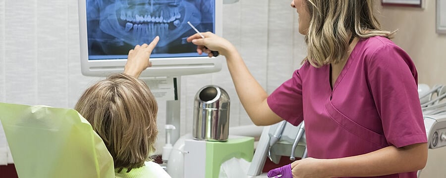
Computer screens used to view clinical images need to be set up, so that they present the images clearly and enable the person reporting on the images to extract all the relevant information. The screen resolution, brightness, and contrast may all need adjusting to achieve this. And the ideal settings for viewing clinical images will not necessarily be the same as the ideal settings for reading text or performing other tasks, such as using the practice management software. Test images such as the TG18QC pattern shown here can be used to configure screens used for reporting on clinical images.
264
They can also be used to carry out routine tests of the screen every three months as described in paragraph 5.3.3 in Appendix I of the Dental GNs. The level of illumination in the room should be set so that the screen can be viewed comfortably. The screen should be positioned so that it can be viewed free from any glare or distracting reflections, such as those from room lights or windows. The results of these checks should be recorded and retained.
293.7
Film-based imaging requires close attention to the film handling and processing conditions to produce good quality radiographs and avoid the necessity for exposures to be repeated. The instructions provided by suppliers of films, intensifying screens used for extraoral radiography, processing solutions, and processing equipment should include the following and all must be adhered to- the expiry date and recommended storage conditions; the processing conditions, i.e. times and temperatures; changing frequency for processing solutions; cleaning instructions for automatic film processes. The employer’s QA programme will need to take these and other matters into account to ensure a consistently high standard of image quality is realised.
344.4
The QA programme may include procedures and records to control film stock, ensure that automatic processes are cleaned and the chemicals are changed regularly, check the light tightness of dark rooms, check the condition of film cassettes and intensifying screens and confirm that they are used with spectrally matched film, ensure that overall QA checks are made on image quality perhaps using a step wedge as shown here.
376.9
These records and checks are explained in more detail in section 5.3.4 of the Dental GNs.

This video covers the quality assurance programme for image processing and viewing equipment.

Each piece of equipment is introduced and the recommended quality assurance checks are described.

A PDF version of these slides is available in the downloads section below.

This article is from the free online

Dental Radiography: Radiation Protection in Dental Practice
Created by
FutureLearn - Learning For Life

Reach your personal and professional goals

Unlock access to hundreds of expert online courses and degrees from top universities and educators to gain accredited qualifications and professional CV-building certificates.

Join over 18 million learners to launch, switch or build upon your career, all at your own pace, across a wide range of topic areas.

Start Learning now