Skip main navigation

New offer! Get 30% off one whole year of Unlimited learning. Subscribe for just £249.99 £174.99. New subscribers only T&Cs apply

Find out more

Practical optimisation of patient doses

Andrew Gulson talks through the ways in which patient doses of radiation are optimised.
0.6
In this step, we’ll look at the methods that operators of dental X-ray equipments can use to optimise patient doses during dental radiography and dental cone-beam CT imaging.
14.4
Optimising patient doses is an important part of dental radiography. If the dose delivered is either too low or too high, the diagnostic quality of the image may be affected- perhaps so much so that the exposure has to be repeated. Even if the image is diagnostically acceptable, a dose that is higher than necessary increases the risk to the patient with no corresponding increase in benefit. Successfully optimising patient doses and consistently producing images of adequate diagnostic quality go hand in hand, and many factors are involved in achieving these goals.
51.8
In this step, we’re going to recap some of the equipment features that assist in the optimisation of patient dose. We will then look at factors that depend on the knowledge and skill of the person who operates the X-ray equipment, such as correct preparation and positioning of the patient, radiographic technique, and the selection of exposure factors. We’ll assume that everything else that is relevant to the radiation protection of the patient is already in place. For instance, appropriate referral criteria have been considered, and the exposure has been justified and authorised. The X-ray equipment and ancillary equipment to be used are of the appropriate type and in good working condition, and the relevant employer’s procedures are being followed.
97.5
Let’s begin, then, by looking at the optimization of patient dose in intraoral radiography.
106
We’ve already mentioned that using rectangular rather than circular collimation means that the patient’s dose is roughly halved with no loss of image quality.
119.9
Rectangular collimator adapters are available for fitting to X-ray sets that are manufactured with circular collimation, as shown in this image. As shown in this picture, you’ll need to use image receptor holders and beam-aiming devices to accurately align the X-ray beam with the image receptor and prevent the radiograph being coned off or clipped. This will require some training and practice before you start using rectangular collimation for the first time. Rectangular collimation should be used routinely for intraoral radiography unless this is not clinically practical. Examples of this might include radiographing patients with additional needs who may have difficulty keeping still, or patients under sedation or general anaesthesia.
168.5
In the previous step, we’ve also mentioned that using a spacer cone with a focus to skin distance of 30 centimetres instead of a 20 centimetre cone can improve image quality while reducing the dose received by the patient. So if using an X-ray set with different cone length options, the longer cone should always be selected. However, care should be taken to select the appropriate exposure settings. The end of the spacer cone should always be positioned as close to the patient as possible, as shown in this image.
200.1
The X-ray beam spreads out with distance, meaning that if the end of the cone is set further back from the patient, a larger area of the patient’s skin will be exposed to the X-ray beam. This will increase the patient dose.
214.9
This picture shows the positioning of the image receptor in relation to the tooth to be imaged and the X-ray beam for the by bisecting angle technique on the left and the paralleling technique on the right. The paralleling technique should always be used in preference to the bisecting angle technique, as it produces better image quality, more reproducible results, and lower patient doses. However, it might be difficult to use the paralleling technique in some situations- for instance, when patients are under general anaesthetic. And in these cases, the use of the bisecting angle technique is acceptable, although appropriate image receptor holders and beam-aiming devices should be used whenever practicable.
262
Use of the bisecting angle technique may involve the thyroid gland being exposed to the main X-ray beam for some projections, such as for the maxillary incisors and maxillary canines, and this should be avoided whenever clinically possible. Let’s look, now, at the effect of the imaging system used on patient dose. This table shows the results of a survey of doses for patients in the UK from intraoral radiographs published by PHE in September 2020. You can see how the choice of imaging system used influences patient doses in general terms. However, there are some useful points to be made that don’t show up in the numbers above. Both types of digital system offer a dose reduction compared with film.
309
Although some phosphor plate systems have effective speeds comparable to speed group D film, these should be avoided. DR systems generally require a slightly lower dose than CR. However, CR systems have a greater latitude than film and DR systems such that both overexposure and underexposure can still result in near-perfect images. Care is required to avoid unnecessary high exposure with both types of digital imaging, as this may not be apparent in the radiographic image due to the software compensating for the higher dose.
347.5
Where film is used, speed group F film should be the first choice, as this is approximately 20% faster than speed group E film. Speed group D film is about half the speed of E-speed film and is associated with the highest patient doses for film-based imaging. They should no longer be used. Self-developing film, which doesn’t feature in these figures, requires higher patient doses to give a satisfactory image and should only be used where it is impractical to use digital imaging or conventional film in rapid processing facilities, such as during surgery, or general anaesthesia, or in a domiciliary care setting.
395.4
The final, and possibly most important, step in optimising patient doses during intraoral radiography is the operator selecting the most appropriate exposure settings. Most modern dental X-ray sets of any type have an array of buttons on the control panel allowing the operator to select the patient size- generally adult or child- the anatomical view required, and the imaging system used, as shown here. The timers of these X-ray sets are generally preprogrammed with sets of exposure times for every combination of patient size and anatomical view so that selecting a suitable exposure setting should be straightforward. Where the operating potential is selectable, higher kVs generally produced lower patient doses.
445.3
The times for each anatomical view and patient type can be scaled up or down to suit the speed of the imaging system being used by altering a setting on the control panel. This setting may be called by several names, such as mode, film density, sensitivity, or similar. The operator should always check that this is correctly set before pushing the exposure button by checking against the guideline exposure protocol. Instructions on how to check and change the preprogrammed setting should be in the operator’s manual. Everything mentioned so far in terms of optimising patient doses during intraoral radiography applies equally to handheld equipment.
490.9
However, a slightly different approach to patient positioning must be taken, as it’s important to keep the X-ray beam approximately horizontal during the exposure so that the operator is fully protected by the backscatter shield. This will need practicing to ensure that good quality radiographs are produced consistently with no unnecessary repeat exposures. Let’s move on now to look at optimisation of patient dose when using panoramic or cephalometric equipment. With panoramic and cephalometric equipment, when it’s not necessary to take an image with a large field of view, the operator should make full use of the various collimation options available– for instance, sectional images of wisdom teeth or TMJ views with panoramic equipment.
541.9
It’s also important to use the appropriate patient size and jaw shape settings, particularly with panoramic equipment, as this matches the size of the focal trough to the patient.
556.4
Correct preparation and positioning of the patient is important. The patient should be asked to remove any spectacles, earrings, other jewellery, dentures, and piercings that could be in the area to be imaged before being positioned in the equipment. Full use of the equipment’s light-beam alignment aids and immobilisation devices should be made when positioning the patient. The exposure time can last anything from 5 to 40 seconds, so it’s particularly important that the patient is made aware of the importance of not moving during the exposure to avoid the need to retake the radiograph.
594.3
The patient should be advised to keep very still, to swallow and push the tongue up into the roof of the mouth, and to close their eyes just before starting the exposure. You can see here that the choice of imaging system has little influence on patient dose received during panoramic exposures. However, with film-based imaging, it’s important that the operator always checks that the intensifying screen is in good condition and loaded with film of the correct spectral sensitivity before taking the radiograph. Here, you can see that, for cephalometric equipment, static DR systems generally produce the highest patient doses, while scanning DR systems generally produce the lowest doses. The same comments as for panoramic systems apply here, too.
646.3
If film/screen systems are used, the operator should check all is in order before proceeding with the radiograph. Finally, let’s look at optimization of patient dose when using dental cone beam CT equipment. Dental cone beam CT systems can deliver the highest patient doses of any type of dental X-ray equipment, and one factor that greatly influences this is the collimation, more usually referred to as the field of view, or FOV. As for any type of X-ray equipment, the field of view used should be as closely matched to the area of interest as possible.
687.1
So for views of a single tooth, such as for placing a single implant, the field of view size selected by the operator should ideally be about 4 centimetres in diameter by 4 centimetres high.
702.6
As well as reducing the dose to the patient, using a smaller field of view means that there is a smaller area that has to be reported on. Dental practises should not take 3D images that are so large that they extend outside the dental anatomy, as they will not have the training needed to report on the whole image. This should only be undertaken by specialist imaging centres that employ staff with specialist qualifications and training, such as clinical radiologists.
739.7
Dental CBCT imaging is a specialist and developing field of dental X-ray imaging about which it is not possible to go into great detail in this course. However, much of what has already been covered for panoramic radiography is also relevant to Optimising patient doses in dental cone beam CT imaging, with a few additional points that are worth mentioning, as shown here. For more information on optimising patient does in dental CBCT imaging, please refer to chapter 4 of the Dental GNs. Just to conclude, successful optimisation of patient dose also depends on other aspects of the overall QA programme, all of which are covered during this course.

This video discusses methods that operators of dental X-ray equipment can use to optimise patient doses of radiation.

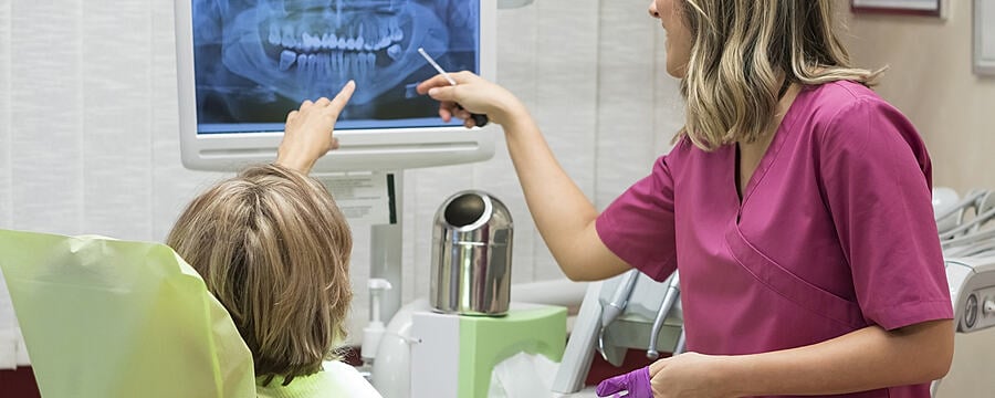
Optimisation of radiation dose relies on specialist features of the X-ray equipment but also on the knowledge and skill of the operators, both of which are discussed in the video. Optimisation features of the different X-ray sets previously covered are reviewed. Considerations for dose optimisation include collimation, focus to skin distance, technique, effect of imaging system, selection of exposure settings and positioning.

A PDF version of these slides is available in the downloads section below.

This article is from the free online

Dental Radiography: Radiation Protection in Dental Practice

Created by
FutureLearn - Learning For Life

Reach your personal and professional goals

Unlock access to hundreds of expert online courses and degrees from top universities and educators to gain accredited qualifications and professional CV-building certificates.

Join over 18 million learners to launch, switch or build upon your career, all at your own pace, across a wide range of topic areas.

Start Learning now