Skip main navigation

New offer! Get 30% off your first 2 months of Unlimited Monthly. Start your subscription for just £29.99 £19.99. New subscribers only. T&Cs apply

Find out more

How does retinopathy of prematurity develop?

An overview of the history of retinopathy of prematurity, how it develops and the changes seen in the back of the eye.
10.8
The retina is the light sensitive tissue which lines the inside of the back of the eye. It is a complex network of photo-receptors and nerve cells (neural tissue). In healthy eyes, when light enters the eye, the retinal cells convert the light energy into electrical impulses which travel up the optic nerve to structures in the brain where the images are interpreted. During foetal development, the eye starts to form very early, about three weeks after conception. Initially development starts as an outgrowth from the brain called the optic vesicle. The different layers of cells in the embryonic eye begin to rapidly divide and develop distinctive types of tissue.
49.2
The outer layers – the lens placode cells – develop into the future lens and the inner layer cells become the future retina.
59.7
By the sixth week, the retina starts to develop from the cells of the optic vesicle. As the future neural retinal cells divide and differentiate, this layer gradually forms the inside lining of the eye behind the lens. This continues to grow into the neural retina and a pigmented layer. A small part of the anterior fifth of retinal layer near the front of the eye further develops into the ciliary body and the iris. Just behind it is the ora serrata. The posterior four fifths continues to develop the photoreceptors and associated neural connections. Throughout this process the eye also continues to grow in diameter and length.
101.7
It is important to note that the neural retina does not have any blood vessels at this stage.
110.8
As the retinal structures continue to differentiate and mature, they begin to require oxygen. This stimulates blood vessels to start to grow outwards from the optic nerve at around 12 to 14 weeks gestation. The development of these new blood vessels is triggered and controlled by growth factors. Some of these are produced by the mother and others are produced in the retina of the baby’s eye. This form is called vascular endothelial growth factor, or VEGF. VEGF is produced in the avascular part of the baby’s retina and this stimulates the growth of blood vessels. These blood vessels spread through the developing layers of the retina as it matures.
148.7
They complete their growth and reach the ora serrata (close to the anterior fifth of the eye) at around normal delivery time (40 weeks) and provide the nutrients and oxygen required for the retina to function. Babies who are born preterm, that is before 37 weeks of gestation, have immature retinal blood vessels that have not yet fully developed and reached the ora serrata. This is a risk for developing retinopathy of prematurity (ROP). After a preterm baby is born, the first sign indicating that ROP is developing is that the retinal blood vessels stop growing outwards.
185.9
This is more likely to happen if the baby has been given too much oxygen in the first few hours, days and weeks of life, or if they have sepsis (widespread infection). This means that the structures of the baby’s retina, which carry on maturing after birth, become increasingly hypoxic (that is not receiving enough oxygen).
208.2
This tissue hypoxia stimulates the production of excessive amounts of VEGF which makes the retinal blood vessels grow abnormally. All the signs of ROP are due to this abnormal proliferation of retinal blood vessels which are fragile, bleed easily and grow in different directions. ROP develops at the boundary between the peripheral avascular retina (which has no blood vessels) and the vascularized retina (which has blood vessels) Fibrous tissues and scarring develop in areas that have bleeding and abnormal new blood vessels. This can lead to further complications such as retinal detachment and resulting in blindness from ROP.
252.3
In summary, the development of the foetal eye and retina is complex and continues right across the whole gestational period. This means that a premature baby born at 28 weeks has a greater risk of having ROP than a premature baby born at 32 weeks. After a preterm birth, early detection and management of ROP can reduce the risk of blindness.

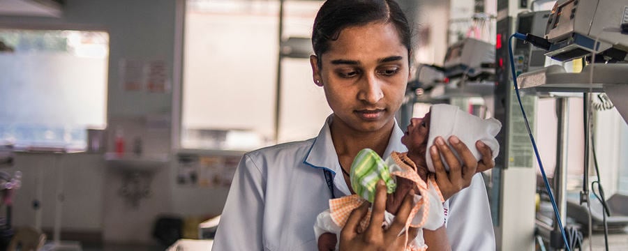
In this video we consider why and how retinopathy of prematurity (ROP) develops and describe the changes that take place in the retina (in the inner layer of the eye). It is important to remember that ROP only develops in infants born four or more weeks premature and that it is only seen in infants who have received neonatal care.

A brief history of ROP

Blindness from ROP was first decribed in the United States of America (USA) in the 1940s, when it was called retrolental fibroplasia (RLF), which means “a fibrous mass behind the lens.” What the ophthalmologists were seeing was a total retinal detachment and scar tissue. At that time the earlier stages i.e., before retinal detachment, had not been described because equipment was not available then to examine the peripheral retina.

In the 1940s and 50s RFL was the single most common cause of blindness in children in high income countries. During this ‘first epidemic’ of ROP the level of neonatal care was poor and ROP related blindness occurred in larger babies with a birthweight of 1000 – 2000g. There was high mortality in the low birth babies.

In 1952 Szewczyk suggested that misuse of 100% supplemental oxygen given to premature babies, which led to hyperopia (high levels of oxygen in the blood) and sudden episodes of anoxia (lack of oxygen), was a cause of RFL. An increase in the rate (incidence) of RFL cases was seen in hospitals that had introduced incubators with high oxygen concentrations for preterm babies.

By the mid-1950s abundant clinical and experimental data showed that RFL was due to overuse of supplemental oxygen. Studies identified the appropriate levels of oxygen preterm babies required and hospitals began introducing new methods to monitor oxygen levels.

By the 1960s and 70s, in high income countries, most babies who developed severe ROP were born weighing less than 1000g. This was called the ‘second epidemic’ of ROP. In the 1990s, treatment in the form of cryotherapy became available for ROP for the first time, and over the last two decades laser has become the standard treatment.

However, the situation has been and remains different in low- and middle-income countries which have been going through a ‘third epidemic’ of blindness due to ROP since the 1980s (Gilbert et al 1997). This third epidemic is a mixture of the first two epidemics and is characterised by high rates of severe ROP developing in both relatively mature and immature preterm babies, reflecting the varying levels of neonatal care provided (Shah et al. 2009). This is compounded by lack of inadequate screening and treatment services. In high-income settings, most infants developing the severe stages of ROP now have extremely low birthweight (less than 1000g) and high quality screening and treatment means that blindness is largely prevented.

The understanding of ROP and its risk factors continue to evolve, which influences practice. In your experience of neonatal care or ROP services, what are the challenges in keeping up to date with evidence based practice?

This article is from the free online

Retinopathy of Prematurity: Practical Approaches to Prevent Blindness

Created by
FutureLearn - Learning For Life

Reach your personal and professional goals

Unlock access to hundreds of expert online courses and degrees from top universities and educators to gain accredited qualifications and professional CV-building certificates.

Join over 18 million learners to launch, switch or build upon your career, all at your own pace, across a wide range of topic areas.

Start Learning now