Skip main navigation

Biology toolbox: Enzyme-substrate interactions and inhibition

How do enzymes interact with their substrates?
Enzyme inhibition.
Cartoon of a flask containing white liquid and producing bubbles
© Setareh Chong

How do enzymes and substrates interact?

There are two models for enzyme-substrate interactions:

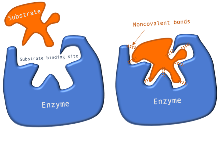
Model 1: Lock and Key In this model, the shape of the active site and substrate complement in such a way that the substrate fits into the binding site perfectly. There’s some truth in the lock and key model in that enzymes do have active sites, which need to be filled with a substrate and interact with the substrate through non-covalent interactions. The problem with this model is that if the substrate fitted the enzyme perfectly, catalysis would be hampered. This is because the enzyme needs to bring about a change to the substrate and not just to bind it. The Lock and Key model explains that the enzyme needs to bind substrate, but once the reaction progresses to the transition state and product formation, the active site would not be able to accommodate this change. This is the paradox of how enzymes work, they need to be able to bind specifically to the substrate, but they also need to be able to turn the substrate into something else (product), which means those two things are at odds with each other. So the enzyme needs to bind the substrate slightly imperfectly in order to be able to turn it over, that is, convert it to the product.

Cartoon illustrating the Lock and Key model of substrate binding which shows two copies of a cartoon enzyme with an opening labelled as the substrate binding site. In the first copy a substrate molecule that is the same shape as the binding site is floating outside the enzyme. In the second copy of the enzyme, the substrate molecule has been fitted in the binding site (like a key in a lock) and is held there by non-covalent bonds Figure 1: A diagram showing the Lock and Key model of enzyme-substrate interaction.

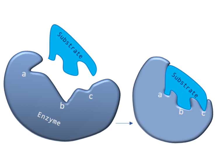
Model 2: Induced Fit In the Induced Fit model, the enzyme active site forms in response to substrate binding. In the diagram, sites a, b and c move in response to the binding substrate. So initially the active site is not perfect, but upon binding, it is able to move, which puts the active site under strain. This strain is then able to elicit the energy that’s required for the reaction to occur by stabilizing the transition state and not just binding of the substrate. The enzyme carries out its work by inducing the substrate to take up a transition state on the path to the required product. In this figure the image on the left shows a cartoon enzyme with a misshapen substrate binding site that has three labelled features (a, b and c). A substrate molecule, which has a different shape to the substrate binding is shown above the enzyme. The image on the right shows the change in shape that occurs in the enzyme as the substrate engages - the labelled features have been "induced" to change shape due to the interactions between the enzyme and substrate. Figure 2: A diagram showing the Induced Fit model of enzyme substrate interaction

Enzyme Inhibition

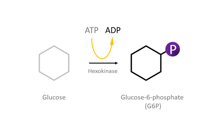
One of the best things about enzymes is that they can be regulated. Regulation is important as this is how a cell controls when a reaction takes place and when it does not. Enzymes are regulated by effector molecules which are most commonly inhibitors. One way of regulating enzymes is by inhibiting them. For example, the breakdown of glucose to pyruvate, glycolysis, is a multi-step process that involves 10 enzymes. The first enzyme in this pathway, hexokinase, which phosphorylates glucose to glucose 6-phosphate (G6P), is inhibited by its own product glucose 6-phosphate (G6P). This way the activity of hexokinase is dampened down to control glycolysis.

A cartoon showing a glucose molecules which is converted to glucose-6-phosphate through the action of the hexokinase enzyme which requires the hydrolysis of ATP Figure 3: Hexokinase converts glucose to glucose 6-phosphate during glycolysis Note: G6P is very structurally similar to glucose (G6P= glucose with an added phosphate group).

There are two types of enzyme inhibition mechanism:

  • Competitive inhibition
  • Non-competitive inhibition

Competitive Inhibition

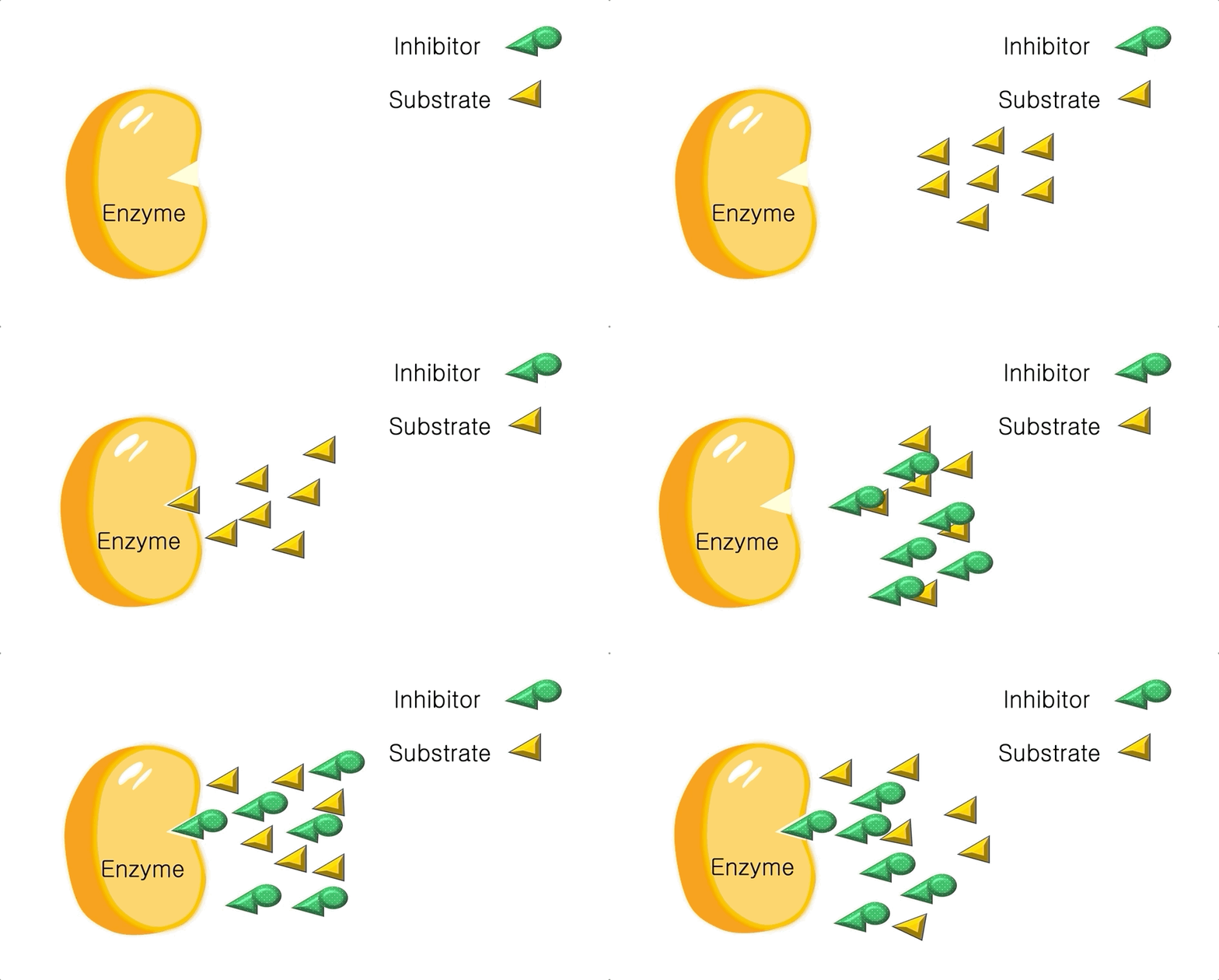
Inhibition by this mechanism involves molecules that are similar to the substrate (substrate analogues) binding to the active site, inhibiting the reaction. They compete with the substrate for binding the enzyme active site. Reaction products that have similar structures to substrates can be used in a feedback loop to inhibit enzyme activity by competing with the substrate for the active binding site. For example G6P (example above) inhibits hexokinase by competing with glucose.

An animation of how competitive inhibition occurs. Substrate molecules are shown binding to the binding site of an enzyme. Then inhibitor molecules appear. These have some features that are the same as the substrate molecules, which allows them to bind to the same binding site as the substrate: the competition between the inhibitor and substrate shows the inhibitor bound and the substrate unable to access the enzyme binding site

Figure 4: Animation of competitive inhibition

Non-competitive Inhibition:

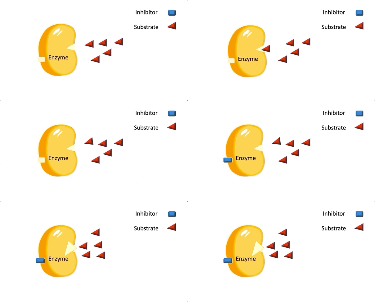
Non-competitive inhibitors don’t compete with substrate for the active site. Instead they bind to a site distant from the active site. This binding effects a conformational change to the structure of the enzyme that changes the shape of the active site in such a way that the active site can no longer accommodate substrate binding. This process of binding at one site to bring about an effect at a distance is called allostery. Non-competitive inhibition is also known as allosteric inhibition.

In the animation of non-competitive inhibition the enzyme is illustrated with two binding sites: one where the substrate can bind, the other where the inhibitor can bind. When the inhibitor binds to its binding site on the enzyme, the substrate binding site changes shape and the substrate is no longer able to bind to the enzyme

Figure 5: Animation of non-competitive inhibition

An example of allosteric inhibition is the inhibition of the phosphofructokinase (PFK) enzyme (also known as the pacemaker for its important role in regulating the rate of glycolysis, thus “setting the pace”), which catalyzes the ATP-dependent phosphorylation of fructose-6-phosphate to synthesise fructose 1,6-bisphosphate and ADP during glycolysis. When the ratio of ATP to ADP is high, ATP binds to the ATP binding site of this enzyme (which is distant to the active site) and inhibits the enzyme. This means that the cell senses that there is too much ATP around so it stops or slows down glycolysis by inhibiting PFK to stop it from making more ATP. This process is reversible. So when the ratio of ATP:AMP is lowered, the enzyme is activated to make more ATP.
© Setareh Chong
This article is from the free online

The Biology of Bugs, Brains, and Beasts

Created by
FutureLearn - Learning For Life

Reach your personal and professional goals

Unlock access to hundreds of expert online courses and degrees from top universities and educators to gain accredited qualifications and professional CV-building certificates.

Join over 18 million learners to launch, switch or build upon your career, all at your own pace, across a wide range of topic areas.

Start Learning now