Skip main navigation

New offer! Get 30% off your first 2 months of Unlimited Monthly. Start your subscription for just £35.99 £24.99. New subscribers only T&Cs apply

Find out more

Function of the heart

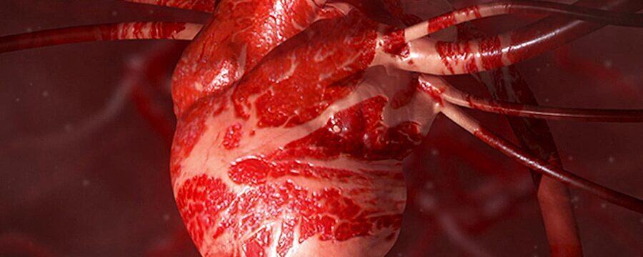
The function on the heart explained by Dr Sam Boateng in a video on the free online course, Heart Health: A Beginner's Guide to Cardiovascular Disease
6.3
We’re going to look at the function of the heart. The main function of the heart is to supply blood to the tissues of the body, delivering oxygen and nutrients to them. Now, the heart is able to function because it is able to generate its own electrical activity. And this starts in the region known as the pacemaker, or the sinoatrial node, that is located in the right atrium. The electrical activity begins in the sinoatrial node, then spreads across the rest of the atrium, through to the other atrium, and then down into the ventricles. This causes the atria to contract before the ventricles.
48.6
Now, the electrical activity of the heart can be measured using the electrocardiogram, or the ECG, that can measure the electrical activity of the heart, and this is depicted by letters of the alphabet P, QRS, and T. Now when the electrical activity of the heart passes through the atria, this causes the atria to contract and, as a result of the polarisation of the atria, this produces a P-wave on the electrocardiogram. It’s important to mention at this point that the atria always contracts before the ventricles and that allows the blood within the atrium to be pumped into the ventricles. And so the atrium must always contract before the ventricles.
95.9
This is facilitated by fibrotic tissue that insulates the electrical activity between the atrium and the ventricles. This means that electrical activity can only pass from the atria to the ventricles through a small region called the atrioventricular node. As the electrical activity passes through the node and into the rest of the ventricles, there is depolarisation of the ventricles which causes them to contract. Now, on the ECG, this produces the QRS complex, which is a large deflection caused by the electrical activity in the ventricles. Following a period of contraction also known as systole - that’s the contraction of the heart - there is a period of relaxation called diastole. This is associated with repolarisation of the ventricles.
152.2
Now, on the ECG, this is depicted by the T-wave that is also a deflection that can be measured, and this is associated with ventricular relaxation or repolarisation of the ventricles. Now, in some cases, a small U-wave is also visible, and this is due to repolarisation of the papillary muscles that are associated with the atrioventricular valves. So it can be seen that these four chambers work together to ensure that the right side pumps deoxygenated blood to the pulmonary circulation to the lungs, whilst on the left side, oxygenated blood is pumped to the systemic circulation.

Join Dr Sam Boateng as he explains the function of the heart and its importance to other systems in the body.

You can download the Week 1 supplement, which contains additional images and descriptions to help you understand the topics covered in this video.

This article is from the free online

Heart Health: A Beginner's Guide to Cardiovascular Disease

Created by
FutureLearn - Learning For Life

Reach your personal and professional goals

Unlock access to hundreds of expert online courses and degrees from top universities and educators to gain accredited qualifications and professional CV-building certificates.

Join over 18 million learners to launch, switch or build upon your career, all at your own pace, across a wide range of topic areas.

Start Learning now