Skip main navigation

New offer! Get 30% off your first 2 months of Unlimited Monthly. Start your subscription for just £29.99 £19.99. New subscribers only. T&Cs apply

Find out more

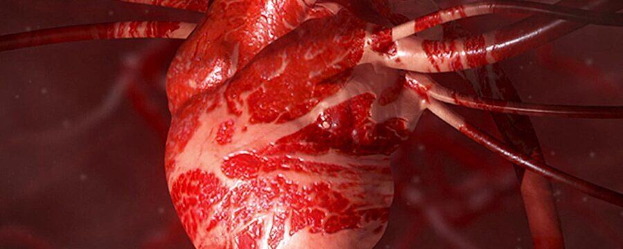
Part 2: Investigating the internal structure of the heart

Heart dissection demonstration to investigate the internal structure of the heart for a Beginner's guide to Heart Health by the University of Reading.
2.5
Now that we’ve looked at the outside of the heart, let’s take a look inside. We’re going to do this by carrying out a longitudinal section through the heart. We’re going to insert a probe into the aorta - that’s the central blood vessel - and then we’re going to cut down the length of it. Please take care to insert a probe and not your finger into the vessel, as if you use your finger as a probe, you’re very likely to cut yourself. So using the probe, insert it.
35.2
I’m going to use a scalpel to cut down the length of the probe, but you can be using a kitchen knife. Keeping your fingers well away, carefully start to cut down the length of the probe, cutting through the tissue all the way down to the apex of the heart. Work your way down the whole of the length of the probe.
62.1
And then carefully remove the probe, and you can open up the heart. We can see here the left ventricle. I’ve also cut through the left atrium. So we’ve opened up the left atrium and the left ventricle. Immediately, you can see the inside of the heart, the heart wall, and we can see these fibrous cords or tendons - the chordae tendineae. Let’s have a look at the right side. We insert the probe into the pulmonary artery, which takes us down into the right ventricle. Let’s cut down the length of the probe. Again, don’t use your finger as you’re likely to cut yourself, cutting down the length of the probe all the way down and then opening out the heart.
122.8
And again, we can see this time the right side to the heart. We can see into the ventricle. We can see the thinner muscle wall. And again, we can see the chordae tendineae, these fibrous cords inside.
139.5
Let’s start by taking a closer look at the heart wall. It’s easier to see on the left-hand side. The heart wall is made of three layers. We’ve got the endocardium, that lines the inside of the heart. Then we have the myocardium, or heart muscle. Surrounding the whole heart, we have the pericardium.
167.5
What we’re going to do is measure the thickness of the heart wall. As we’re scientists, we’re going to measure in three different places. So take your ruler and measure the thickness of the left ventricle in three different places.
190.2
And then note these on your worksheet.
198.2
Let’s do the same on the right-hand side. Opening the heart out, our much thinner heart wall. Let’s measure its thickness in three places and note those on the worksheet.
227
Let’s take a moment to think about the function of those heart walls. Blood enters the left atrium, travels into the left ventricle, and from there, it’s squeezed out through the aorta to travel all the way around the body. On the right-hand side, blood enters the right atrium, travels into the right ventricle, and from there, it’s squeezed out through the pulmonary artery only as far as the lungs. Therefore, the muscle on the left-hand side has to work much harder. It needs to be much bigger, much thicker, much stronger to push the blood all the way around the body than the muscle on the right-hand side.
282
Let’s look at the atrioventricular valves now. The atrioventricular valves lie between the atria and the ventricles on each side of the heart. They’re there to prevent the flow of blood in the wrong direction. Blood enters through the atrium, travelling into the ventricle. The ventricle then squeezes or contracts, forcing the blood out through the aorta, and at the same time, it forces the atrioventricular valves closed.
316.4
But these valves are forced so fast that they’re at risk of being turned inside out like an umbrella on a windy day. If we look inside the heart, we can see these fibrous chords, the chordae tendineae. And the muscles that they’re attached to, these are the papillary muscles. When the heart contracts, the papillary muscles also contract, tensing through these cords and holding the valve closed so it can’t be blown inside out.
353.7
On this left-hand side of the heart, the heart valve, atrioventricular valve, is known as the mitral or bicuspid valve. That’s because there are two cusps or two flaps to the valve. We can see here two papillary muscles, one here and one here, attached to two groups of chordae tendineae. There’s one group here, if you imagine that was closed around, and there’s another group around the back. So two papillary muscles, two groups of chordae tendineae come in to two cusps.
396.6
On the right-hand side, if we thread our probe through the right atrium, we can see it emerging into the right ventricle, coming out through the atrioventricular valve. On the right-hand side, the atrioventricular valve is known as the tricuspid valve because it has three cusps. There are also three papillary muscles - we can see one here; the other two are tucked away - attaching to three groups of chordae tendineae, which then attach to the valves. We can see the valve on this left-hand side here. The valve looks quite delicate, but it actually has to be quite strong to withstand the forces of the blood.
452.3
As well as the two atrioventricular valves, there are also the semilunar valves, and these lie between the ventricles and the major vessels, so between the left ventricle and the aorta and between the right ventricle and the pulmonary artery. They’re less easy to see, particularly when the vessels have been cut through. But you might be able to see them on your specimen.
479.1
That concludes our investigation of the outside and the inside of the heart. Complete your worksheet and then dispose of all your waste appropriately. You need to wash all of the utensils with hot, soapy water and wipe the surface down with an appropriate disinfectant. Finally, wash your hands thoroughly. I hope that you’ve enjoyed this practical and found it both interesting and helpful in improving your understanding of the structure the heart. Remember, you can share and discuss your findings online.

In this video we look at the internal structure of the heart. You should still have everything set up from the previous step. If you haven’t, make sure you’ve got everything set up as it is listed.

There will be a discussion in the next step where you can share your thoughts about the exercise.

Dr Natasha Barrett and the University of Reading are happy for these videos to be used as learning resources in teaching. If you do wish to use the home practical videos, please ensure that you also include the safety information which accompany each practical. The safety video for this practical is in Step 1.13 and in the article in Step 1.14. If you wish to use the video or any other video on the course, please attribute the University of Reading. Please do not modify any of the videos from the course.

This article is from the free online

Heart Health: A Beginner's Guide to Cardiovascular Disease

Created by
FutureLearn - Learning For Life

Reach your personal and professional goals

Unlock access to hundreds of expert online courses and degrees from top universities and educators to gain accredited qualifications and professional CV-building certificates.

Join over 18 million learners to launch, switch or build upon your career, all at your own pace, across a wide range of topic areas.

Start Learning now