
Classification of microbes

All life can be divided into Prokaryotes and Eukaryotes. The table below shows the differences between these two groups:
| Prokaryotes | Eukaryotes |
|---|---|
| More simple | More complex |
| Prone to mutation | Resistant to mutation |
| Circular chromosomes anchored to cell wall | Chromosomes in the nucleus |
| Virulence plasmids | No extra DNA elements |
| No organelles | Cellular organelles |
| Prokaryotic ribosomes | Eukaryotic ribosomes |
| Small size and simplicity allows rapid growth | Slow cell division |
We will now discuss the classification of bacteria.
Between bacteria, many variances exist and can be used to assist with the classification of bacteria. This includes their shape, the way they cluster into groups, the type of cell wall (Gram staining), metabolic processes, and resistance to antibacterials.
Shape
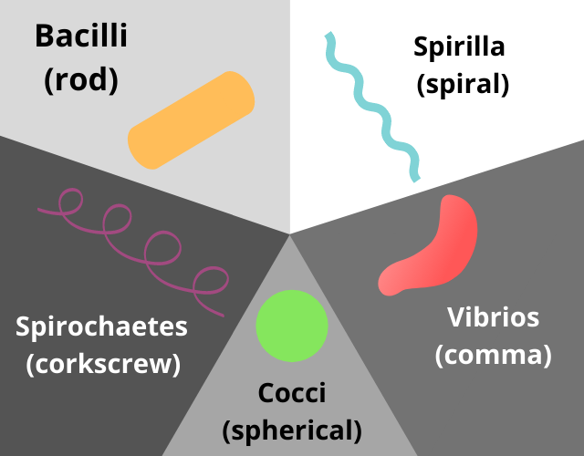
One of the ways to classify bacteria is by shape. The diagram above details the differences in bacterial shape, and what these look like.
Groups
The way in which bacteria arrange themselves into groups can be used in classification. These groups may be clusters, such as Staphlococcus aureus: 
Or they may be ‘beads on a string’, such as Streptococcus pyogenes: 
Cell wall
The Gram Staining method was developed at the end of the 19th century to distinguish one bacterium from another, based on the chemical and physical properties of their cell walls. This includes detecting the presence or absence of peptidoglycan in the bacterial outer layer, and the stain colour reveals this information. The Gram-staining technique is carried out through the sequential use of 2 stains – first a crystal violet (purple), followed by safranin (dark pink). Gram-positive bacteria retain the purple stain, whereas Gram-negative do not. To watch a video of a gram stain being performed in the lab, click here
| Gram-positive | Gram-negative |
|---|---|
| Presence of peptidoglycan layer | Absence of peptidoglycan layer |
| Stains purple | Stains pink-red |
If you would like to learn more about Gram-negative bacteria, you may be interested in the FutureLearn course Challenges in Antibiotic Resistance: Gram Negative Bacteria.
Some examples of organisms that fall into each category are below:
| Gram- positive | Gram- negative | |
|---|---|---|
| Coccus | Staphylococcus aureus, streptococcus pyogenes | Neisseria meningitidis, Neisseria gonorrhoeae |
| Bacilli | Corynebacterium diphtheriae, Clostridium perfringens | Escherichia coli, Pseudomonas aeruginosa |
Metabolic Processes
Oxygen use: Aerobes vs Anaerobes
Aerobic bacteria require oxygen to grow.
Anaerobic bacteria grow in the absence of free oxygen.
Bacteria can be classified into four groups according to their requirement for oxygen:
- Obligate (strict) anaerobes – grow only in the absence of oxygen
- Facultative anaerobes – grow in the presence or absence of oxygen
- Microaerophilic – grow best in low oxygen concentrations
- Obligate aerobes – grow only in the presence of oxygen
Catalase
The presence of the enzyme catalase can be used to differentiate between streptococci and staphylococci – staphylococci are catalase positive.
Haemolysis
The ability of bacteria to lyse red blood cells (haemolysis) or convert haemoglobin to a green pigment on a culture plate can be used to classify bacteria.
- Alpha-haemolytic bacteria – no lyses but does convert haemoglobin to a green pigment
- Beta-haemolytic bacteria – lyse RBCs
- Gamma-haemolytic bacteria – do not lyse RBCs
Now we have explored a variety of techniques to classify bacteria, the next step will provide an example of how these may be used in practice.
Let us know in the comments below: do you use any of the above techniques in your setting? Are there any different methods of classifying bacteria you are aware of?


Reach your personal and professional goals
Unlock access to hundreds of expert online courses and degrees from top universities and educators to gain accredited qualifications and professional CV-building certificates.
Join over 18 million learners to launch, switch or build upon your career, all at your own pace, across a wide range of topic areas.


