Skip main navigation

New offer! Get 30% off one whole year of Unlimited learning. Subscribe for just £249.99 £174.99. New subscribers only T&Cs apply

Find out more

Troubleshooting and Making Changes to Invasive Ventilation

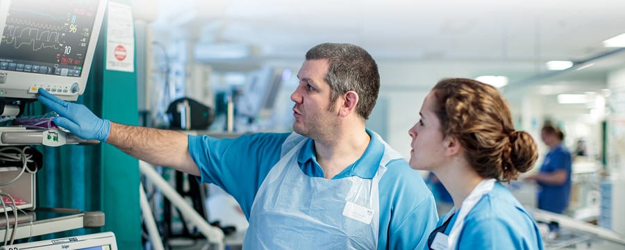
Troubleshooting and Making Changes to Invasive Ventilation video
1.3
Hello. My name is Duncan Birse, and I’m one of the Intensive Care Registrars here in Edinburgh. Welcome to this tutorial. Today, we’re going to have a look at how to make changes or adjustments to invasive ventilation for patients in critical care. The first thing to say is that, hopefully, you’re already familiar with the main modes of invasive ventilation. If you’ve not done so already, why don’t you have a look at the ventilation tutorial, which explains these modes in more detail? Once you’ve done that, have a go at the simulation exercise where you’re asked to set up a ventilator for two different patients.
40.8
Another important point is that there are lots of different makes and models of ventilator. The exact terminology or the acronyms used for different modes of ventilation vary between brands. However, the basic concepts remain the same. It’s important that you’ve received adequate training and are familiar with the ventilators that you use in your hospital.
64.6
Let’s have a look at the ventilator screen. Again, these can vary a lot between different ventilators, but here is a typical setup. The ventilator mode is displayed often in more than one place. The numbers along the bottom represent the settings that we can control and adjust for this mode of ventilation. In this case, we’re using a pressure control mode, called SIMV, which stands for synchronised intermittent mandatory ventilation. So we can control the fraction of inspired oxygen, the FIO2, the inspiratory pressure, the inspiratory time or TI, the frequency of breaths or the respiratory rate, the positive end-expiratory pressure, known as the PEEP, and the pressure support should the patient take a spontaneous breath.
115.4
One setting you might be used to using is the I:E ratio, or inspiratory to expiratory ratio.
121.9
As this is a time cycled mode, the I:E ratio is determined by the inspiratory time. If you want to make adjustments, selecting the TI will usually
131.8
display the I:E ratio and allow you to make changes.
138.2
The numbers on the right-hand side are the values which the ventilator is actually recording. Given that this is a pressure control mode, the tidal volume delivered is determined by the compliance of the patient’s lungs and chest wall. So we can see how changes to the inspiratory pressure affect the tidal volume by looking at this value over here on the right. The other numbers help demonstrate whether the settings we have set are actually being delivered to the patient. The middle of the screen contains three graphs, plotting pressure, flow, and volume against time. These can provide some very useful information, but we won’t talk any more about these just now. Sometimes, flow-volume loops are displayed in this portion of the screen.
185.2
Finally, these very small numbers represent our alarm limits. So if the recorded values fall below or rise above these limits, an audible alarm will sound, and a visible alarm will display on the screen. The setup of these screens is customizable. And so again, it’s really important that you are familiar with how your ventilators display the same information.
210.8
The way in which you change the settings, again, depends on the ventilator you’re using. Many modern ventilators have touchscreens, although some older versions or transport ventilators have a set of buttons which allow you to access the menus. Once you’ve chosen the desired setting, you usually increase or decrease it by rotating a knob. In order to confirm the change, you need to press the knob. This is important, otherwise the ventilator won’t actually change the setting.
241.8
So let’s get started making some changes to patient ventilation. There are six scenarios coming up. Try and work out what changes you would make to the patient’s ventilation settings yourself. We’ll then give you our suggestions. There will be more than one right answer for each scenario, but these examples will highlight some important principles. First up, we’ve got a 64-year-old man. He weighs 87 kilogrammes. He has contracted COVID-19. And he’s on his second day of invasive ventilation here in the ICU. You notice that his peripheral saturations have dropped to 82%, and you perform an arterial blood gas. The main feature on this blood gas is his degree of hypoxia.
293.2
So he has a PaO2, arterial partial pressure of oxygen, of only 6.8 kilopascals. A chest X-ray performed earlier in the day, as reported by the radiologist, is showing left basal atelectasis. And here are his current ventilation settings. So he has an FiO2 of 40, an inspiratory pressure of 24, an respiratory rate of 24, and a PEEP of 4.
326.7
Here’s all the information for this scenario summarised on one page. Have a look at it and work out what changes you think you would make to his ventilation. The first thing to say, actually, the first thing you might do is disconnect the patient from the ventilator and bag them using 100% oxygen to allow their sats to come up. Let’s assume you’ve done this, and you’re wanting to put the patient back on the ventilator. What changes would you make to prevent further desaturations? You’ll notice a little 30-second timer has appeared in the top right-hand corner. Take this time to write down the changes that you would make, and then we’ll show you our suggestions shortly.
368.8
If you need more time, feel free to pause the video and take as long as you need.
405.1
So how did you get on? Hopefully, you’ve written some settings down. Let’s have a look at our suggestions. So this patient’s main problem was hypoxia. So let’s start by increasing the FiO2, say, from 40% to 60%. The hypoxia seemed to be mainly caused by atelectasis, or collapse, particularly in the left lower zone, as seen on the chest X-ray. So at the moment, the PEEP is only 4. This is relatively low. In order to help recruit this left lower zone and in order to prevent further atelectasis from developing, let’s increase the PEEP from 4 to 10. This is going to splint open the alveoli and the small airways, and hopefully, prevent further atelectasis from forming.
456.3
What’s really important is that whatever changes you make to the ventilation, you go back and assess the impact of these changes. Some effects you’ll be able to see straight away. So we’ll be continuously monitoring this patient’s peripheral saturations, and we might see a change immediately. Other changes take longer to see. And for example, we’re going to want to repeat an arterial blood gas in 30 minutes’ time to assess the impact of our changes, providing that the patient’s peripheral sats haven’t dropped in the meantime. OK, let’s move onto our next case. This is a 35-year-old man who weighs 92 kilogrammes. He suffered a traumatic brain injury, which has resulted in a diffuse axonal injury.
506
He’s on his second day in intensive care, having been intubated and ventilated in the accident and emergency department for reduced conscious level. And he has an intracranial pressure monitor in situ and an ICP of 24 millimetres of mercury at present. And here’s his blood gas. So the main feature of this blood gas is hypercarbia. He has a PaCO2, a partial pressure carbon dioxide, of 6.2 kilopascals. This is slightly higher than the normal range, but it’s definitely higher than we would want for a patient such as this. In a patient with a raised intracranial pressure, cerebral vasodilation caused by hypercarbia can be particularly detrimental and can cause a rise in ICP.
556.8
We would want to target a PaCO2 of between 4.5 and 5.0 for this patient. So here’s his current ventilation. Similar to our first patient, it’s a pressure control mode, SIMV again, except this time with the addition of VG. This stands for volume guarantee. This means that we can set a tidal volume, and the ventilator will deliver it as a pressure-controlled breath using the minimum pressure required. So our patient has an FiO2 of 30%, a tidal volume of 550 mils, and a respiratory rate of 18.
598.3
Here’s all the information summarised on one page. Have a think about the changes that you are going to want to make. Again, the 30-second timer has appeared in the top right corner. Feel free to pause the video if you think you need more time.
645
Very good. Hopefully, you’ve got some new settings written down. Let’s have a look at what we thought. So in order to reduce this patient’s PaCO2, we’re going to want to increase the minute ventilation. The minute ventilation is determined by tidal volume and respiratory rate. This patient already has a relatively large tidal volume for their body weight, particularly for their ideal body weight, and so we probably wouldn’t want to increase this any further because of the risk of inducing a ventilator-associated lung injury. Instead, in order to increase the minute ventilation, we can increase the respiratory rate, say, from 18 to 22.
683.6
Again, we’re going to want to assess the impact of this change, and we can do this by monitoring the patient’s end-tidal carbon dioxide, but also by repeating an arterial blood gas in about half an hour’s time to check that their PaCO2 has come down to within our target range.
702.3
All right, let’s move onto our third patient, which is a 56-year-old woman who weighs 55 kilogrammes. She has developed a streptococcal pneumonia, for which she’s required invasive ventilation on intensive care. This is her 12th day on the intensive care unit, and she’s currently weaning from invasive ventilation. The nurse looking after the patient has come to speak to you because they’re worried about the tidal volumes. In particular, the ventilator is alarming, saying the tidal volumes are high. Here is a blood gas from the patient.
747.1
The ventilator settings are as follows. So you can see the patient is on a fully spontaneous mode. This ventilator calls this mode CPAP pressure support. And if you look at the flow-volume graph, you can see that the colour of the waveform has changed from a blue to a grey. This, again, indicates that these are spontaneous, or patient-triggered, breaths. The patient’s receiving 40% oxygen with a PEEP of 7. And for each of these spontaneous breaths, a pressure support of 15. This is creating a tidal volume of over 600 mils, which is greater than our upper alarm limit of 600, and is causing our ventilator to alarm. So here’s all the information for this patient.
792.8
Take 30 seconds, again, to write down some settings. Feel free to pause the video if you need a bit longer.
830.5
Excellent. So hopefully, you’ve got something written down. Let’s have a look at what we thought. So the problem here is that the pressure support being provided to the patient is too much. They’re weaning from ventilation, they’re getting stronger, and they don’t need as much pressure support. So this additional pressure support is generating excessively large tidal volumes.
854.2
We want to try and limit tidal volumes during invasive ventilation to around 6 to 8 mils per kilo. This has been shown to reduce the risk of ventilator-associated lung injury. And for this patient at 55 kilogrammes, 6 mils per kilo would be around 330 mils. So the first change we’re going to make is we’re going to reduce the pressure support, say, from 15 down to 10. You should very quickly be able to see the effect of this on the tidal volumes and continue titrating or adjusting the pressure support as required until the tidal volumes are appropriate for this patient.
897.3
So let’s have a look at our fourth patient. This is a 44-year-old man. He weighs 92 kilogrammes, but has an ideal body weight of 75 kilogrammes based on his height. He has ARDS– secondary to gallstone pancreatitis. And the nurse looking after the patient has come to speak to you because they’re concerned about the peak pressures. In fact, the ventilator is alarming.
924.4
Here’s a blood gas. You can see the patient is slightly acidotic, and a slightly high partial pressure carbon dioxide at 7.4. Here are the ventilator settings. So this time, the patient is on a volume control mode, volume-controlled SIMV, with 40% inspired oxygen, tidal volume’s at 600, respiratory rate of 18, and a PEEP of 12. And as you can see, the peak inspiratory pressure is 32. This is higher than our upper alarm limit of 30, and so the ventilator is alarming.
967.2
So here’s all the information displayed on one page. Take 30 seconds, again, to write down some changes you would make to this patient’s ventilation. This is a slightly more complex case, so if you need a bit longer, again, feel free to pause the presentation.
1014.9
So what did we think for this case? Well, the main problem here is the peak pressure of 32. This is higher than our alarm limit and higher than we would like for most patients in intensive care, where we try to limit peak pressures, and in particular, plateau pressures, to less than 30. So what can we do about this? Well, the tidal volume at the moment is set to 600. And the patient’s ideal body weight is 75 kilogrammes. So based on 6 mils per kilo, the patient should be receiving a tidal volume of around 450. So the first thing we can do is reduce that tidal volume, say, to around 4.75.
1059.1
The next thing is that the inspiratory time is only 0.6 seconds. This is relatively short, and for the patient’s respiratory rate will mean that there’s a very short inspiratory time compared to the expiratory time.
1075
What this means is that the same volume is delivered over a shorter period of time, resulting in higher pressures. So by increasing the inspiratory time, we can also hopefully reduce the peak pressures. So, say, increasing the inspiratory time from 0.6 to 0.8. And then finally, the patient is receiving 12 of PEEP. They’re only on 40% oxygen, and their oxygenation was good on the arterial blood gas, so they probably don’t need as much PEEP as this. By reducing the PEEP, say, from 12 to 10, we will also reduce the peak pressures. So there’s a number of different things we can do here to help limit peak pressure. We can ensure our tidal volumes are set appropriately.
1123.4
We can increase the inspiratory time so that the tidal volume is delivered over a longer period of time on the mandatory breaths, therefore reducing the peak pressure.
1136.3
And if appropriate, we can also reduce the PEEP.
1143.2
Right, last couple of patients. Let’s have a look at our fifth patient. This is a 53-year-old woman. She weighs 78 kilogrammes. She also has COVID-19. She’s on the 19th day of her intensive care stay. She’s had a tracheostomy inserted and is currently weaning from ventilation. And she looks uncomfortable. Her breathing pattern is very awkward, and she doesn’t look settled on the ventilator. Here is an arterial blood gas for this patient, which, despite her abnormal breathing pattern, actually looks very normal. Here are her ventilation settings. So she’s on pressure control, SIMV.
1195
And we can see from the waveforms that she’s receiving a mixture of mandatory breaths, and in between these, taking her own spontaneous breaths. The settings down on the bottom say that she’s got an inspiratory pressure of 18 for the mandatory breaths and a pressure support of 8 for the spontaneous breaths. This is on top of a PEEP of 8 and a mandatory respiratory rate of 20. So here’s all the information summarised, again, on one page. You’ll be getting used to this by now. So take 30 seconds to write down some new ventilation settings. Press pause if you need a little bit longer.
1265.6
Excellent. Well, let’s have a look at what we thought. So the main problem here seems to be that the patient is not synchronising correctly with the ventilator. So although this is a synchronised intermittent mandatory mode of ventilation, on occasions, there’s not appropriate synchronisation with the ventilator. And we can see this because if we look at the pressure and flow graphs, there are patient-triggered, or spontaneous, breaths stacked on top of mandatory delivered breaths. Now there are several ways in which we can deal with this problem. The first one might be that we want to sedate the patient and/or muscle relax them. But this is a patient who is weaning from ventilation, and so this isn’t going to be an appropriate step.
1317.9
Something else we could consider doing is reducing the respiratory rate, say, from 20 to 12. This is going to reduce the mandatory rate that the ventilator delivers and allow more time in between the mandatory breaths for the patient to take their own spontaneous breaths. This might help with synchronisation with the ventilator. An alternative might be to change the mode of the ventilator altogether to a purely spontaneous mode. It may be that the patient is strong enough now to wean onto a spontaneous mode and doesn’t require any additional mandatory breaths or a backup ventilatory rate.
1361.6
And here’s our final patient. So this is a 26-year-old woman. She weighs 69 kilogrammes. She’s been admitted with a severe exacerbation of asthma and is currently desaturating. The ventilator’s alarming because her peak pressures are very high. And here’s a blood gas. So she has a respiratory acidosis with a PCO2 of 7.8 and a PO2 of 8.
1394.3
And here’s the ventilator settings. So she’s on a volume control mode. Her FiO2 is 40%. Her tidal volume is 400. Respiratory rate’s 24. And PEEP is 10. And the ventilator’s alarming because the peak inspiratory pressure is 35.
1416.8
So here’s all that information on one page. Your 30 seconds has already started. Write down some new settings. And if you need a bit longer, then pause the video.
1448.4
Great. So let’s see what we got from this one.
1455.8
This patient is an asthmatic. And asthma is a disease of the small airways that results in difficulty, particularly, in exhalation. We can see this from our ventilator, particularly if we look at the flow-time graph. And if we look at the end of the expiratory phase here, we can see that the next inspiratory phase starts before all of the gas has been fully exhaled because the flow has not reached 0. As a result, gas is trapped within the alveoli, and the lungs are continuously expanded, resulting in hyperinflation. This can be very problematic and make patients difficult to ventilate. So there are a number of things we can do to help this patient.
1504.7
The first one is we can reduce the inspiratory time. This might seem slightly counter-intuitive, given that we’ve already got high peak pressures. But what this is going to do is it’s going to allow a longer time for expiration, and allow all of that gas to leave the lung, and hopefully, reduce the amount of gas-trapping. Along with reducing the inspiratory time, we’re also going to reduce the respiratory rate.
1532.8
And this combination is going to significantly increase our I:E ratio, so increase the amount of expiratory time compared to inspiratory time. Again, all of this is going to help gas leave the lungs during the expiratory phase and reduce the amount of gas-trapping. And to help with our peak inspiratory pressures, we’re going to reduce our PEEP. We’re actually going to take our PEEP from 10 down to 0. Severe asthma is one of the few cases where we might consider using a PEEP of 0 in intensive care, particularly because the patients have such narrow airways that they actually generate a significant amount of what’s called auto-PEEP.
1578.6
So although we’re setting a PEEP of 0, the end expiratory pressure within the lungs will never actually reach 0.
1587.3
So the combination of increasing the I:E ratio by reducing the inspiratory time and the respiratory rate, and dropping our PEEP to 0, will hopefully help reduce our peak inspiratory pressures and also prevent further gas-trapping. Again, we’re going to assess these changes. Some things you’ll be able to see straight away, such as changes in the flow-time curve. Other things you may need to assess in a half an hour’s time with a repeat arterial blood gas. We can probably tolerate what we call permissive hypercarbia for this patient, i.e., we can allow their partial pressure of carbon dioxide to rise, and we can allow a degree of respiratory acidosis, providing they are otherwise cardiovascularly stable.
1639.9
So how did you get on with those six scenarios? If you came up with different suggestions to us, why not post them in the comments below, along with your reasons why? Maybe you have different equipment. Maybe you find other strategies work better. We’d be really interested to know were there any differences in the ways patients are ventilated in different parts of the world. Thank you very much for listening, and I hope you enjoy the rest of this course.

This video demonstrates how to manage ventilation in the critically ill patient by working through patient scenarios.

This article is from the free online

COVID-19 Critical Care: Understanding and Application

Created by
FutureLearn - Learning For Life

Reach your personal and professional goals

Unlock access to hundreds of expert online courses and degrees from top universities and educators to gain accredited qualifications and professional CV-building certificates.

Join over 18 million learners to launch, switch or build upon your career, all at your own pace, across a wide range of topic areas.

Start Learning now