Skip main navigation

New offer! Get 30% off your first 2 months of Unlimited Monthly. Start your subscription for just £29.99 £19.99. New subscribers only. T&Cs apply

Find out more

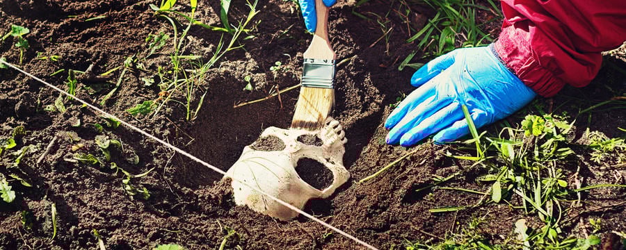
Патология скелета и травматология

Выявление патологии и травм - важный этап судебно-медицинской экспертизы.
Доктор Ребекка Гоуленд дает пояснения по этой теме.
9.3
In this section we’re going to talk about skeletal pathology, so abnormal changes on the bones. As you might expect, this is a vast subject, there’s been lots of textbooks written on it. And we teach a whole Master’s Course on skeletal pathology here at Durham. From a bioarchaeological perspective, we look at skeletal pathology to try and find out about how people lived in the past, how they interacted with each other and their environment. From a forensic perspective, we look at skeletal pathology in order to try and identify the individual, so we look at certain features that might help to identify them.
44.6
So for example, dental disease, if they have a healed fracture that they sustained as a child, if there’s any evidence of surgery, we can compare those features to ante-mortem records or medical records, and it can be confirmed with discussions with the family. We also look at pathology to find evidence of peri-mortem trauma or traumatic lesions that might be consistent with the cause of death, or might tell us something about the circumstances of death. There are a wide variety of diseases that can affect the skeleton. But the skeleton has a limited response to disease. It either takes bone away, forms new bone or a combination of both.
89
So what I’m going to do here is just take you through some of the basics of recording these changes, so that later on, you can perhaps attempt a diagnosis. Starting with new bone formation, bone will react to inflammation or trauma by forming new bone. And this is in the form of this woven bone, which as you can see, is a slightly different, darker gray colour from the normal bone surface. It’s mechanically weak, and it’s quite porous as well. And when we see this woven bone, we know that the disease process or the inflammation was active at the time of death. As the bone starts to heal, this woven bone becomes remodeled and forms what we call lamellar bone.
126.6
And you can see here, you’ve got this striated bone on the surface, it’s the same color as normal bone, and it’s a similar texture. And this shows us that the bone is healing at the time of death.
151.4
On this bone here, you can see that the lamellar bone is very irregular, and the bone actually feels really heavy to touch as well. And there’s been a lot of new bone formation and remodeling. And it’s created this irregular appearance. It’s not just affecting the surface of the bone, but it’s actually affecting the cortex of the bone too. And it can even affect the bone marrow. When you’re recording new bone formation, you want to record whether it’s woven bone, or lamellar bone or a combination of both. You want to record the appearance of it, is it porous? Is it pitted? Is it striated? Is it irregular?
189.7
And you want to record the location on the bone, where the new bone formation is. Is it diffuse across the whole length of the bone? Is it localized just in a patch of bone? And is that on the proximal or the distal surface of the bone? As well as bone formation, you can get bone destruction. So if you see here on this cranium, if you ignore these patches of post mortem damage, and focus instead on these lesions, you can see that there are different areas of destruction. Now the edges of some of these lesions are remodeled. So that tells us that there’s been a bony response to that destruction.
231.1
In some disease processes, such as secondary metastatic carcinoma, where cancer has spread from, for example, the liver and spread to the bones, you’ll see that the edges of the lesions are very sharp and ragged, which indicates that it’s a very fast acting disease process. It’s important when you’re recording bone destruction to record the size of the lesion, the location of the lesion, but also to note the margins of the lesion. Has there been a bony response to that disease process, or are the edges very sharp. You should also record whether it’s a single isolated lesion, or whether there are multiple lesions as well.
273.8
If you look at the distribution of lesions throughout the skeleton, and that goes for new bone formation as well, that can help you diagnose a specific disease, because different diseases will have different distributions throughout the body. Diseases can also have an impact on the shape of bone because they’re affecting the quality of bone produced. One disease is vitamin D deficiency, otherwise known as rickets in children. So I’ve got two femora here. You can see on this side, it’s a normal shape, but this one is quite sharply bowed. And this is because vitamin D is needed for the normal mineralisation of the bones.
311.5
So when there’s not enough of it, the bone that it’s produced is mechanically quite weak and can easily become deformed. Even in an adult skeleton, we can still see signs of stress during childhood. One of the signs of stress is known as enamel hypoplasia. When the teeth are forming during childhood, and the enamel is being laid down, if the child is malnourished in some way or is ill, it will disrupt this enamel formation, and it can produce pits or lines on the teeth. Another childhood condition that we see quite commonly, is known as cribra orbitalia, and that refers to these lesions in the orbit or eye socket.
353.8
What you’re seeing here is an expansion of the marrow cavity (diploë) within the bone and a thinning of the outer table. And so it looks like this porosity is on the eye socket. This is a childhood disease, but these lesions can still be retained into adulthood. So if we look at this adult here, you can see this slight porosity here. And that’s evidence of healed, a healed childhood episode of cribra orbitalia. One of the most common skeletal pathologies is joint disease. Here is an example. And you can see these bony spurs around the margins of this vertebra and that’s to do with intervertebral disc degeneration.
396.3
So as the disc degenerates, the body produces these spurs to try and help prop the vertebrae up mechanically. If we look at the underneath of this vertebra, as well, you can see this indentation here. This is called a schmorl’s node. And again, this is associated with the degeneration of the intervertebral disc. If we look at these vertebrae here, these are the neck vertebrae, you can see that the osteophytes, those bone spurs, have become so big that the vertebrae have actually fused together in a solid block. These are the vertebra from the neck and so this would clearly have restricted movement in the neck. It could of caused the individual pain, but this is very difficult to interpret from the skeleton.
444
But if it had affected that person during life, then it could become a useful identifying feature for the forensic anthropologist. Another example of joint disease is on this femur here. So you can see the head of the femur, which is at the hip socket, that there’s massive new bone formation all around the margins of the joint. And you could see this would have actually changed the margin of the joint. You can see quite a large spur underneath here. Now we know from this bone that it would have affected the individual’s gait, so the way that they walked, because we can see changes to the muscle attachment sites as well.
479.4
So that might have been a useful identifying feature for this person. Finally, we can see on this humerus, although it’s quite subtle, you can see this shiny patch of bone and some slight porosity here. This is osteoarthritis. So the joint cartilage has worn away, and the bone ends have been rubbing against each other. And that’s what creates this shiny surface. And again, while this may not have affected the individual too much during life, there may be some inflammation in the medical records that can help establish their identity. Healed fractures to the bones can also be useful for identifying an individual because they can quite often be linked to medical records.
515.4
So here we have the tibia and fibula from a single individual. And you can see that there’s a fracture here to the proximal fibula, and through here, towards the distal end of the tibia. And this is quite well healed. So it’s a fracture that occurred some time ago. Unfortunately, we’re not able to say exactly when it happened in this person’s life, but we can certainly see that the individual has lived quite some time with it. Here’s another example of a healed fracture. So here, we’ve got the fracture to the proximal femur.
556.5
And you can see the shortened neck of the femur here, and all this new bone growth here, which is essentially the results of the soft tissue damage that occurred alongside the fracture. But again, on this individual, it’s well healed. There’s also no real sign of joint disease to the head of the femur, which you sometimes get after a fracture because it alters the mechanics of the joint. Forensic odontology, the recording of the teeth and dental disease, is really important for identifying individuals within forensic contexts. While not everyone will have dental records, most of us do end up going to the dentist at some point in our lives.
595.3
And if we can match these records, to the remains of the individuals within a forensic context, then that’s really useful for establishing identity. Dental pathologies can be really useful. So for example, caries, which are these holes that appear in the tooth crowns, usually associated with eating too much sugar. Quite often we go to the dentist now to have these filled. You can see overcrowding of the teeth here at the front of this jaw. Quite often we would get braces or some kind of dental intervention there. And this can be useful for identifying the individual. So these can be matched to formal dental records. But sometimes, particular dental morphologies can cause an unusual appearance or a distinctive appearance.
649.4
So anecdotal evidence from families or friends about the person’s appearance, whether they had a gap in their teeth, whether their teeth were crowded or rotated could still be useful alongside formal dental records. So here, we’re going to go through some different examples of trauma. When you’re recording trauma, it’s really important to know whether it’s ante-mortem, so whether it occurred prior to death, whether it’s peri-mortem, so occurring at around the time of death, or whether it’s post-mortem, occurring after death. So I’ve got some different examples here for us to take a look at. So here we’ve got an example of ante-mortem trauma. You can see at some point in this individual’s life, they sustained quite a large injury to their skull.
689.1
And you can see it’s quite lengthy, but it’s very well healed. So it looks like it might have been caused from sharp force trauma, so such as a blade, but either way, it’s very well healed. And it’s very difficult to make out the margins of that injury. So for this individual here, this injury could easily look like a post-mortem break, because you can see that the edges are quite white and quite ragged. But actually, you can see that there’s been some new bone formation here. So this is an ante-mortem fracture, and the body has had some time to try and knit the two fractured ends together. And we can see new bone formation around those fractured edges.
727.6
But then the individual died before it could completely heal. This individual here has sustained some sharp force trauma. And this looks to be peri-mortem. So we can see that the fracture edges are very similar in color to the bone. You can also see some shininess and some striations, which indicate that this trauma was inflicted, when the bone was still green, i.e. still fresh. In this example, you can see that the entire top of the cranium has been removed. If you look at the edges of the cut, you can see that there is similar colour to the bone and they also have that smooth, shiny surface. So they’re very similar to the peri-mortem cuts that we’ve just seen.
782.8
Now, thankfully, these actually occurred post-mortem. And this is part of a post-mortem procedure called a craniotomy, where the top of the skull is removed. Because the cuts were done on bone that was still fresh, you can see that they still have the same features as peri-mortem trauma. And so it can be quite difficult to differentiate between these two forms of trauma. But we know, as you can see, that this is an anatomical specimen or medical model, you can see by these hooks and pins. So it was used for display, so we know that this occurred post-mortem. On this skull here, if you look at this hole, it’s quite regular and round and neat.
820.3
And so it could be mistaken for peri-mortem trauma. But actually, if you look at the edges of the lesion, you see that they’re very white, and they’re quite sharp. So that indicates that this was inflicted after the bone was dry. So this is post-mortem. So we’ve shown you some examples of sharp force trauma. So here I’m just going to show you some examples of healed and peri-mortem blunt force trauma. Firstly, we’ve got an example of ante-mortem blunt force trauma. It’s quite hard to spot but there are two injuries, there and there. They’re two depression fractures that are well healed.
869.2
If we compare those to this individual here, where you can see that the injury is peri-mortem, there’s no evidence of any healing here. And again, it’s caused through blunt force trauma. This is the skull of a child and your eye is naturally drawn to this hole here. But what I want you to focus on first is this line here. This indicates that there was actually a fracture at some point here, so there was some trauma there. And this is concentric fracture line that occurred around that point. This break here, is actually a radiating fracture line. So when you have blunt force trauma, you often get radiating and concentric fracture lines coming from it.
908.8
This hole here is also peri-mortem, but this is actually an attempt by a doctor to trepan the child to release the pressure on the brain caused by the injury. Unfortunately, the procedure was unsuccessful, and the child died because there is no sign of healing on any of the bones. This is a scapula. And the injury that I want to show you here is this. This is a stab wound. So it’s created this puncture through the bone, and you can see that it’s come out the other side and you can see that flake of bone coming away. This section has provided a basic introduction to some of the pathological conditions that you’ll see most commonly in skeletal remains.
964.9
We’ve also shown you different examples of trauma, and how to differentiate between anti-, peri- and post-mortem trauma. There’s a whole raft of literature out there for you to take a look at. Obviously, we’ve just covered the basics here. But please do have a look at the reading list attached. We’ve now come to the end of this series of instructional videos. Our aim is for you to use these alongside the text, images, and interactive 3D models that we’ve provided on the online course. Together these resources will help you to learn the key techniques for humans skeletal analysis. So thank you for watching. We hope that you found these videos useful.
1006.9
If you’re interested in furthering your knowledge in this area, then please either look at our online course or take a look at the Durham University website to see the other courses that we offer. Thank you!

В данном разделе мы обсудим патологии скелета, то есть аномальные изменения костей. Как вы, наверное, догадываетесь, это обширная тема, которой посвящено множество учебников, а здесь, в Даремском университете, мы ведем целый магистерский курс по патологиям скелета.

С биоархеологической точки зрения мы рассматриваем патологии скелета для того, чтобы выяснить, как люди жили раньше, как они взаимодействовали друг с другом и с окружавшей их средой. С судебно-медицинской же точки зрения патологии скелета изучаются для целей установления личности. Кроме того, патологические изменения могут говорить о травмах, нанесенных в момент наступления смерти, или о травматических повреждениях, указывающих на причину смерти, а также быть источником информации об обстоятельствах смерти.

Существует множество заболеваний, которые могут отразиться на скелете, однако набор реакций костной ткани на заболевания ограничен. Происходит либо разрушение костной ткани, либо образование новой костной ткани, либо два этих процесса вместе. Итак, сейчас мы с вами пройдем кое-какие основы фиксации этих изменений, чтобы позднее вы, возможно, попробовали провести диагностику.

Примечание и дополнение к переводу: термин биоархеология впервые был введен британским археологом Грэхемом Кларком в 1972 г., как ссылка на зооархеологию или исследование костей животных из археологических памятников; в других странах используются так же названия остеоархеология или палео-остеология; по сравнению с биоархеологией, остеоархеология – это научное исследование, которое сосредоточено исключительно на человеческом скелете; фактически процесс раскопок, эксгумации и исследования останков человека являются различными стадиями биоархеологических исследований.

This article is from the free online

Forensic Archaeology and Anthropology (Russian)

Created by
FutureLearn - Learning For Life

Reach your personal and professional goals

Unlock access to hundreds of expert online courses and degrees from top universities and educators to gain accredited qualifications and professional CV-building certificates.

Join over 18 million learners to launch, switch or build upon your career, all at your own pace, across a wide range of topic areas.

Start Learning now