
New Bone Formation and Destruction

Although bone is a plastic and responsive tissue, it is limited in the ways in which it can respond to pathological or traumatic insults. Bone can form new bone or it can destroy existing bone tissues. Because these responses are non-specific, multiple pathological conditions can produce similar outcomes. It is important to carefully analyse and record any signs of pathology and even small details can be significant for correctly identifying a pathological condition.
New Bone Formation
New bone formation is relatively slow and can take approximately 10 times longer than bone destruction. New bone formation is usually classified into three categories; woven, lamellar or a mix of the two types. Identifying the type of new bone can be useful in understanding the timing of a particular pathological condition or disease process present in an individual.
Woven Bone
When new bone is first produced it has a distinctive porous appearance and is usually a slightly different colour (grey/brown) in comparison to mature bone. It is referred to as ‘woven bone’. This new bone is weak, grey/brown in colour, and it often has a pitted or striated surface. Woven bone can be produced in response to trauma or inflammation. If woven bone is present on the skeleton it means that the disease process or trauma was recent or active at the time of death. For example, if woven bone is visible around the margins of bone fracture then it indicates that the individual had lived for at least one to two weeks after the fracture and prior to death. The features of woven bone are as follows:
- Mechanically weak, random collagen fabric, porous
- Grey/brown in colour, usually pitted or striated
- Indicates that the disease process was active at the time of death
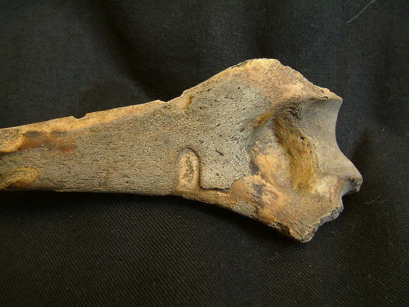 This bone demonstrates a thick layer of very distinct woven bone. Woven bone can be very subtle and may not be as clear as in this image.
Lamellar Bone
Over time, woven bone remodels, developing a more regular and mechanically stronger structure to form lamellar bone. Lamellar bone is the same colour as ‘normal’ mature bone because this is essentially what it is. However, the appearance of lamellar bone is often striated or pitted. In some instances, it can look very irregular and this is often the case if it is produced as a result of a chronic, or long term pathological process that has affected the bone. Lamellar bone is typically:
- Mechanically strong, regular collagen fabric
- Same colour as surrounding ‘normal’ bone, but often pitted, striated or irregular in morphology
- Indicates that the disease process was healing at the time of death
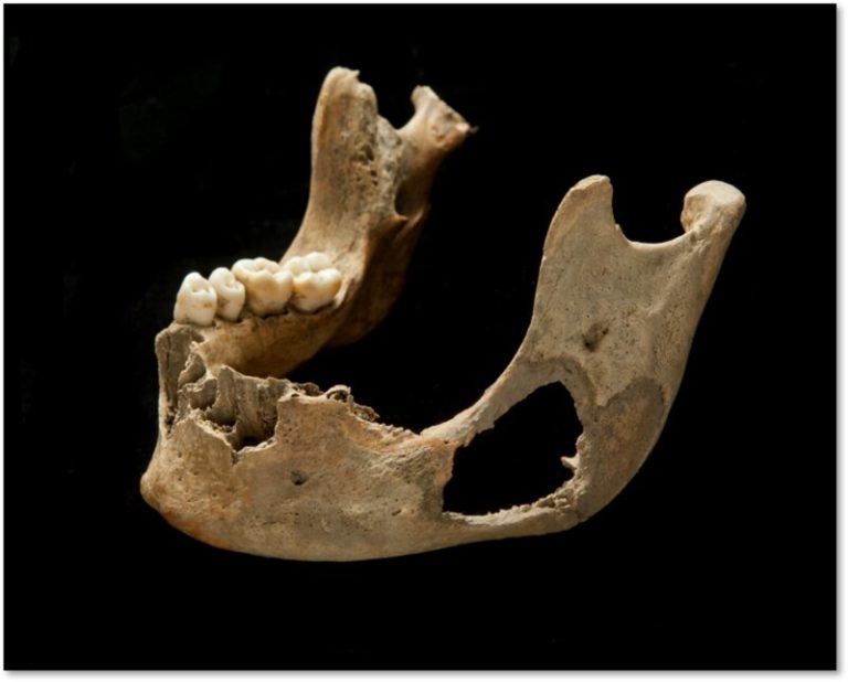
 An example of lamellar bone formation. Note the striated appearance and colour.
 These two tibiae show extensive lamellar and woven new bone formation, indicating a longstanding infection that was also still active at the time of death.
Bone Destruction
Bone destruction can either involve localised or generalised bone loss, depending on the type of disease.
General Bone Loss
Many of us are familiar with the condition osteoporosis, which many older people suffer from, as a general loss in bone mass. This bone loss leaves the skeleton mechanically weaker and can lead to fractures.
If a person undergoes a period of starvation, this can also result in a general loss of bone mass. This is because the bones act as a mineral store for various functions of the body and in the absence of food these gradually become depleted. Individuals suffering from anorexia or enduring forced starvation will, therefore, have weaker bones due to the loss of bone mineral.
Localised Bone Loss
Bone destruction can occur in single or multiple locations on either one or multiple bones across the skeleton. The appearance, the location of these lesions on the bone, and the distribution across the skeleton are all important factors for diagnosis. When recording pathological lesions, it is important to note the size of the lesion(s), the precise location, and the appearance of the margins of the lesion. Bone destruction can result from dietary imbalances, infectious disease, neoplastic disease (cancer), joint diseases and hormonal imbalances.
Very aggressive diseases can destroy bone rapidly. In such instances, the margins of the lesion will be sharp and irregular, with no evidence of new bone formation in response to the destruction. If a pathological condition is long-lived (chronic), then the affected bone will respond by forming new bone and the edges of the destructive lesion will gradually become remodelled. In some diseases, the edges of the lesion are surrounded by rounded and dense bone. This indicates some healing of the lesion.
 Bone destruction with sharp edges and some bone response in the form of woven bone around the jaw. This indicates an aggressive condition, but one that was chronic enough for the bone to respond. This is from our published research, Roberts et al. 2016, International Journal of Palaeopathology, after the link click ‘download pdf’. You may also be interested in this short piece in The Conversation on childhood malnutrition.
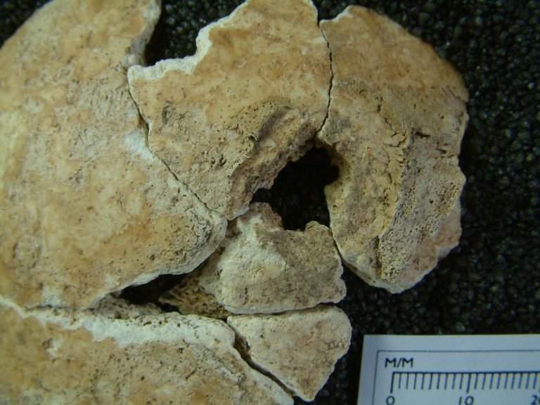 Destructive lesions in the cranium surrounded by some new woven bone. Again this indicates an aggressive condition, but one in which the bone has had time to respond and therefore longstanding. Other lesions in this skeleton are at different stages of remodelling. This is possible tuberculosis in a child. This case is from our published research and open access is available here: Gowland et al. 2018, Bioarchaeology International.
Edges of Lesions
The speed with which bone is destroyed may be important for diagnosis. Irregular, sharp margins with no evidence of bone formation indicate rapid destruction. Margins that are smooth or sclerotic indicate some bone formation (healing).
Recognising pathological lesions can be complicated! There’s a lot to remember and consider. Practice identifying the features of pathological bone with the 3D models provided in the ‘SEE ALSO’ section below. Then continue to explore this fascinating topic by moving on to the next article focused on recording pathological lesions.

Reach your personal and professional goals
Unlock access to hundreds of expert online courses and degrees from top universities and educators to gain accredited qualifications and professional CV-building certificates.
Join over 18 million learners to launch, switch or build upon your career, all at your own pace, across a wide range of topic areas.


