Skip main navigation

New offer! Get 30% off your first 2 months of Unlimited Monthly. Start your subscription for just £35.99 £24.99. New subscribers only T&Cs apply

Find out more

X-ray equipment

The design features, safety and warning features, operating parameters and equipment selection of X-ray equipment used in dental practices.
7.4
In this step, I’m going to look at the design features, safety warning systems, and operating parameters associated with the four different types of dental X-ray equipment. I will also be showing you how these contribute to restricting doses to patients.
23.9
Firstly, we’ll look at the features that are common to all dental X-ray equipment, and then we’ll look at each type of X-ray set in turn. Equipment should be designed to ensure that the levels of leakage radiation do not exceed certain limits. Leakage radiation is X-radiation that passes through the body of the X-ray head, rather than the emission in the useful beam. All X-ray heads have some permanently fitted filtration in the form of aluminium or copper that is used to absorb the lower energy X-rays in the radiation beam, and therefore reduce the dose to the patient, as explained in week one. The amount of filtration in an X-ray set should be stated on a label fixed to the X-ray head.
66
It is expressed in terms of millimetres of aluminium or sometimes millimetres of copper. Let’s look at the safety and warning systems. All dental X-ray equipment should be provided with automatic warning devices, which firstly indicate when the tube is ready to emit radiation. In most cases, this is a power supply light, or the display panel being illuminated. Secondly, it should have a clearly distinguishable signal indicating when X-rays are being emitted. This is usually a light accompanied by an audible warning. The exposure controls should ensure that the exposure terminates automatically after a predetermined condition has been reached, such as when the set exposure time has elapsed. This should also terminate the exposure immediately if they are released partway through the exposure.
115.5
Sometimes this feature is not practical on equipment where the exposure is started from a computer keyboard. And so an emergency stop button, or power isolated button, must be positioned close to the operator to be able to terminate the exposure in an emergency. Turning now to some of the design features that you will see on intraoral X-ray equipment. The focus to skin distance is usually 200 or 300 millimetres. This is the distance from the point of origin of the X-ray beam inside the X-ray tube to the end of the spacer/director cone. Having a longer distance between the X-ray tube and the patient gives a better quality radiographic image, whilst helping reduce dose to the patient.
154
You will note that the cone isn’t actually cone-shaped. They used to be cone-shaped many years ago, but an open ended tube is now used. And this gives it a more uniform X-ray beam. The X-ray beam is shaped, or collimated, to give a fixed X-ray beam size. The shape of the beam should be rectangular and measure approximately 35 by 45 millimetres. The X-ray equipment predominately used to have a circular beam shape, which was required to be less than 60 millimetres in diameter, but the rectangular shape should now be used. Rectangular collimators should be able to be rotated to allow the X-ray beam to be correctly orientated and aligned with the image receptor in the patient’s mouth.
196.3
The image receptor is normally independent of the X-ray set and is usually positioned so that it is in the main X-ray beam by the use of an image receptor holder and alignment tool. The operating potential should be in the range of 60 to 70 kV, with most equipment either having a single fixed value or two settings - usually 60 and 70 kV. A few X-ray sets have a range of settings available. The tube current is usually in the range of 4 to 10 mA, and like operating potential, has either a fixed value or several available settings. The length of time that the X-rays are omitted, the exposure time, is a fraction of a second.
236.6
And times are either adjusted manually by the operator or by the operator selecting a tooth image, corresponding to the tooth being radiographed. This type of exposure timer is called an anatomical timer and has predetermined exposure times already programmed according to the size of the tooth being imaged and the size of the patient. Anatomical timers also have a range of density or sensitivity settings available to allow for the different sets of exposure times required for different types of imaging systems. Usually, only a single type of imaging system is in use in a practise. And so this density, or sensitivity setting, is only rarely altered.
279.4
In addition to the above, handheld intraoral X-ray sets should have a backscatter shield to protect the operator from x-rays scattered from the patient, some means of preventing X-rays being generated if the unit is left unattended - such as a pin code or a time-out feature that locks the controls after a preset time with no activity - and the ability to quickly switch off the unit at the control panel or remove the battery pack without the operator having to expose their hands to the X-ray beam in the event of an accident. Before I move on to talk about panoramic, cephalometric, and cone beam CT X-ray sets, I’d like to describe some of the features that they have in common.
322.1
They’re all used for extra-oral radiography. That is, the imaging device is used outside the mouth. Direct radiography is predominantly used. The operating potential is manually selected and is usually in the range of 60 to 90 kV, with some cone beam CT equipment going as high as 120 kV. The tube current is selected by the operator, usually in the range of 2 to 12 mA. Exposure times are somewhere between 5 and 20 seconds. The size and shape of the X-ray beam can be changed depending on the type of equipment, the patient being radiographed, and the area or volume being radiographed as well.
361.9
A lot of equipment has preset exposure settings for different patient sizes - children and small, medium, and large adults - which give pre-programmed values of operating potential, tube current, exposure time, and may also change the collimation. There are features on the equipment to allow the patient to be positioned correctly, including light beams, bite pegs, and ear or head locators, with some cone beam CT equipment carrying out a scout exposure so that the operator can confirm the positioning is satisfactory prior to carrying out a full exposure. Let’s look at some of the design features that you may see on panoramic X-ray equipment.
401.1
The distance between the focal spot and the imaging system device is approximately 50 centimetres, with the patient standing at about the midpoint. The X-ray beam is a narrow vertical beam approximately 0.5 centimetres wide and 15 centimetres high at the image receptor. This beam scans around the patient and results in a final radiograph approximately 30 centimetres wide by 15 centimetres high. During the exposure, the equipment will automatically change its rotation speed, or kV and mA settings, to compensate for the increased thickness of the patient’s spinal column while the X-ray beam is directly behind the patient’s head. Turning to some of the uses and design features that you may see on cephalometric X-ray equipment.
449.3
The two most commonly used images are the lateral - side view - or the postero-anterior - back to front view. The distance between the focal spots and the imaging device is normally between 1.5 and 2 metres. The aim of this is to get near parallel X-rays resulting in as little enlargement of the image from the true size, to allow for accurate measurements to be taken from the resulting radiograph. The scanning X-ray beam size for this equipment is approximately 5 to 10 millimetres wide and 24 centimetres high at the detector. Resulting in a final radiograph approximately 18 centimetres wide by 24 centimetres high. There are also cephalometric X-ray sets using direct digital imaging.
495.4
But instead of a vertical X-ray beam scanning, a single short exposure, usually less than one second, is used. In this case, the X-ray beam is the full 18 by 24 centimetres in size at the detector. This is very much like the way that films are exposed and older types of cephalometric X-ray sets. For lateral cephalometry, a soft tissue filter - which is usually additional aluminium filtration - may be positioned over the front section of the image to enable the profile of the soft tissue overlying the patient’s skull and teeth to be imaged. Finally, let’s look at the design features that you may see on cone beam CT X-ray equipment.
534.1
Like a panoramic X-ray set, the distance between the focal spot and the imaging system or device is approximately 50 centimetres, with the patient standing at about the midpoint. X-ray beams are rectangular shaped, and are collimated such that the size of the image volume is around 4 or 5 centimetres in diameter by 4 or 5 centimetres high when used for views of individual teeth such as single implants. For the full upper and lower jaw images required for full implant replacements, a more specialist work, larger volumes may be needed. Actual beam sizes are usually a little larger than the final imaged volumes.
574.7
Most equipment has the option of changing the exposure settings to alter the resolution of the final image, depending on the detail required. The higher resolution images can result in patient doses being significantly higher than the standard resolution or low dose settings.

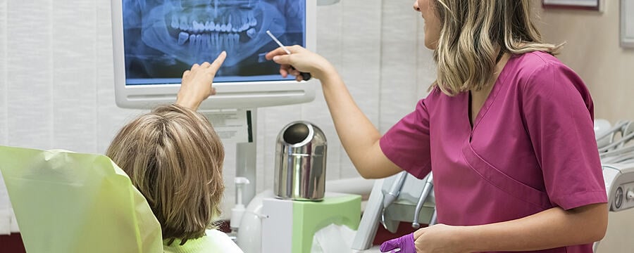
In this video, Graham Ramsden covers key features of X-ray equipment used in dental practices.

This video looks at design features, safety and warning systems and operating parameters of the X-ray equipment previously discussed. The features common to all dental X-ray sets are first covered, followed by the unique features of each X-ray set. The video reviews how these features contribute to optimal function and how they restrict doses to patients.

This article is from the free online

Dental Radiography: Radiation Protection in Dental Practice
Created by
FutureLearn - Learning For Life

Reach your personal and professional goals

Unlock access to hundreds of expert online courses and degrees from top universities and educators to gain accredited qualifications and professional CV-building certificates.

Join over 18 million learners to launch, switch or build upon your career, all at your own pace, across a wide range of topic areas.

Start Learning now