
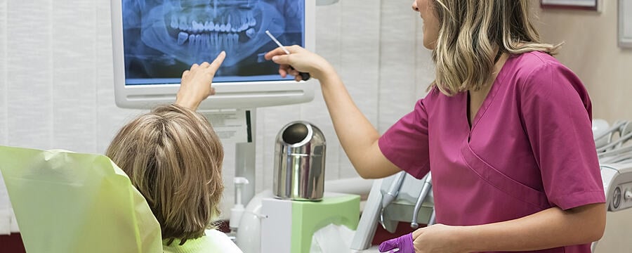
Patient Contact Shielding in Radiography
Patient contact shielding is available for use in radiography – protective lead aprons and thyroid shields (or guards). Both these items incorporate a thin sheet of material (usually lead) to absorb the X-radiation thereby reducing the radiation dose to the patient.
Protective Lead Aprons
Recent publications by the British Institute of Radiology and the dental guidance notes, do not recommend the use of lead aprons for any type of dental radiography.
Following the merger of the FGDP with the College of General Dentistry (CGDent), the Guidance Notes for dental practitioners on the safe use of X-ray equipment (GNs), which are referred to extensively throughout this step have been moved to the CGDent website. If you cannot access the links in this step that take you directly to specific pages in the GNs, please access the Dental GNs here.
As shown below for a typical intra-oral radiograph, dental X-rays are not directed towards the patient’s torso, so the use of a lead apron is of little benefit to the patient and is not recommended, including for patients who are, or maybe, pregnant. Some X-rays may be scattered towards the patient’s torso, but this will be a negligible amount.

Image adapted from Rothband, with permission.
The image above highlights the path of the primary X-ray beam. Note that this does not pass through the torso, so the lead apron worn by the patient is not of any benefit.
Thyroid Shields
The thyroid gland is particularly sensitive to radiation and so thyroid shields have been designed to provide additional protection to the patient’s thyroid gland. A typical shield is shown in the image below. However, there are only a few, limited, situations in dental radiography where the use of a thyroid shield is recommended.

Image sourced from Rothband, with permission.
For intra-oral, panoramic and cephalometric radiography, the use of appropriate equipment, exposure factors, technique (e.g. paralleling technique for intraoral radiography), collimation, or field limitation techniques all provide an equal or greater reduction in the dose to the thyroid gland than the use of a thyroid shield. As these factors also reduce both the effective dose to the patient and the exposure of the operator, these measures should be used in preference to a thyroid shield.
Thyroid shields are recommended for radiography only when the thyroid gland may be unavoidably in the primary X-ray beam and only after seeking the advice of your MPE. Some examples of such situations could be:
- Dental CBCT, panoramic or cephalometric examinations where the field of view extends well below the mandible
- Upper standard (or anterior) occlusal and maxillary central incisor views taken with the bisecting angle technique
- Where a patient requires the use of an unusual positioning technique
When using a thyroid shield for extra-oral imaging it should be ensured that the thyroid shield is not within the primary X-ray beam (so far as it is possible to exclude it), in order to minimise any artefacts caused by the shield that might render the image diagnostically unacceptable resulting in the need for repeat exposure. The image below shows an example of an artefact caused by a thyroid shield in a panoramic radiograph.
Dental Radiography: Radiation Protection in Dental Practice

Dental Radiography: Radiation Protection in Dental Practice
Reach your personal and professional goals
Unlock access to hundreds of expert online courses and degrees from top universities and educators to gain accredited qualifications and professional CV-building certificates.
Join over 18 million learners to launch, switch or build upon your career, all at your own pace, across a wide range of topic areas.


